
| PathWhiz ID | Pathway | Meta Data |
|---|---|---|
PW129307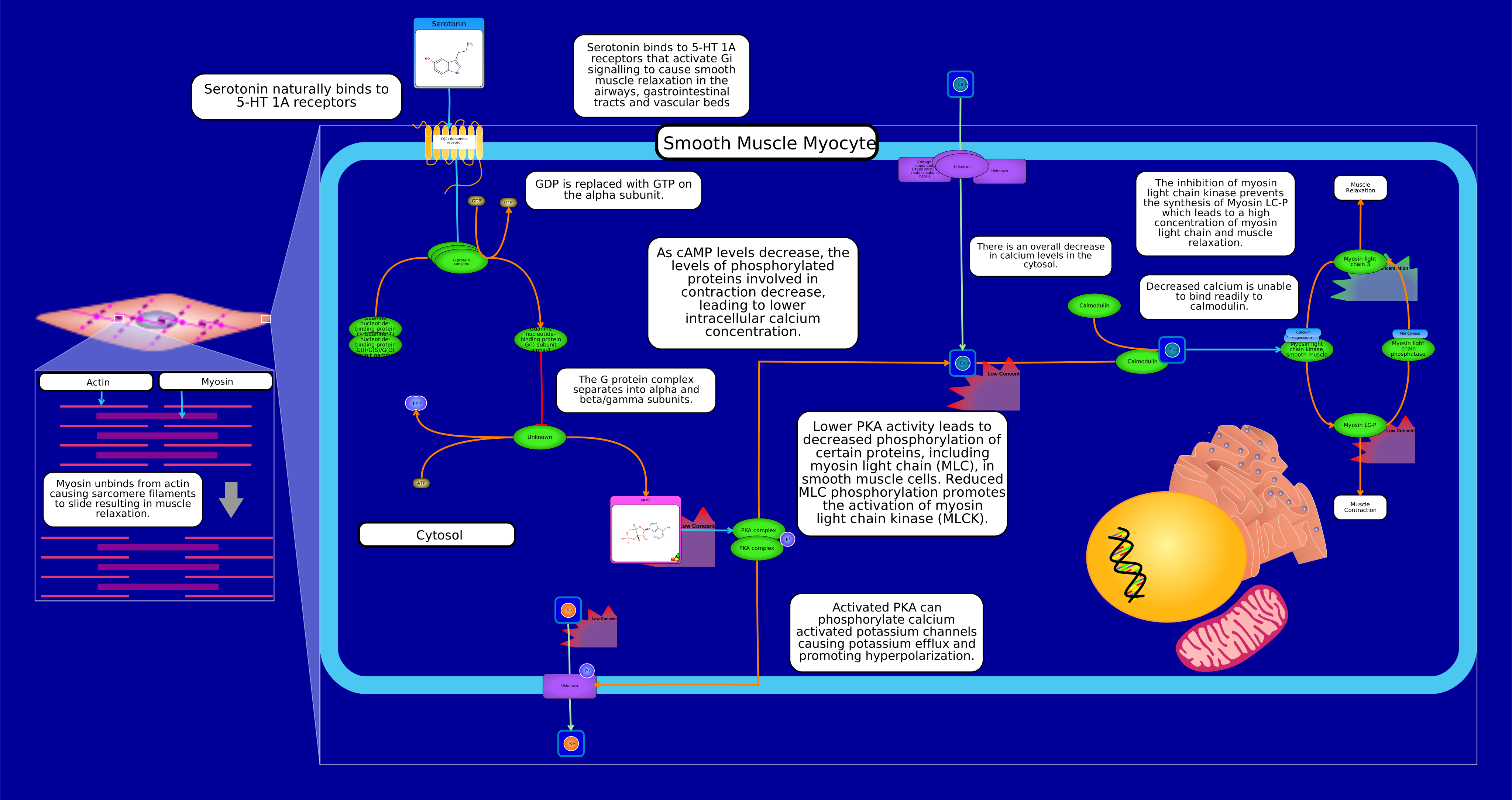 View Pathway
|
physiological
Gi Serotonergic Smooth Muscle RelaxationBos taurus
Although numerous GPCRs have the ability to couple to more than one heterotrimeric G protein, a given GPCR is typically classified based on the G protein subfamily (e.g., Gs, Gi/o, or Gq/11) it preferentially activates. Activation of the 5-HT1A receptor typically leads to Gi protein-mediated signaling, which is commonly associated with smooth muscle relaxation rather than contraction. When serotonin (5-HT) or other ligands bind to the 5-HT1A receptor, it triggers a cascade of intracellular events through Gi protein activation. The Gi protein inhibits the activity of adenylate cyclase, which reduces the production of cyclic adenosine monophosphate (cAMP). Reduced cAMP levels can lead to the deactivation of protein kinase A (PKA) and the inhibition of various downstream signaling pathways. In the context of smooth muscle, this reduction in cAMP levels generally leads to smooth muscle relaxation. The physiological effects of serotonin can vary depending on the specific receptors involved, the tissues in question, and the overall context of the signaling pathways. Different serotonin receptors can have opposing effects on smooth muscle, leading to either contraction or relaxation, depending on the receptor subtype and downstream signaling pathways involved. Serotonin increases the motility of the GI tract muscles, induces muscle constriction in the lungs and uterus, influences vessel muscles in both directions (constriction/relaxation), takes part in platelet aggregation, excites nociceptive pain neurons, and influences CNS neurons. Serotonin also plays a role in the symptoms of GI inflammation, acting through different mechanisms to exert pro- or anti-inflammatory activity.
Summarily, Gi inhibits adenylate cyclase 5 which results in the reduced conversion of ATP to cAMP and reduced activation of Protein Kinase A (PKA). Lower PKA activity leads to decreased phosphorylation of certain proteins, including myosin light chain (MLC), in smooth muscle cells. Reduced MLC phosphorylation promotes the activation of myosin light chain kinase (MLCK). Activated PKA can phosphorylate calcium activated potassium channels causing potassium efflux and promoting hyperpolarization. Low potassium levels, and the resulting hyperpolarization, can affect the activity of voltage-gated calcium channels. These channels are involved in calcium influx, which can, in turn, influence cAMP levels and PKA activity. Calcium ions can activate adenylate cyclase, leading to increased cAMP production and PKA activation.
|
Creator: Omolola Created On: September 14, 2023 at 10:33 Last Updated: September 14, 2023 at 10:33 |
PW128603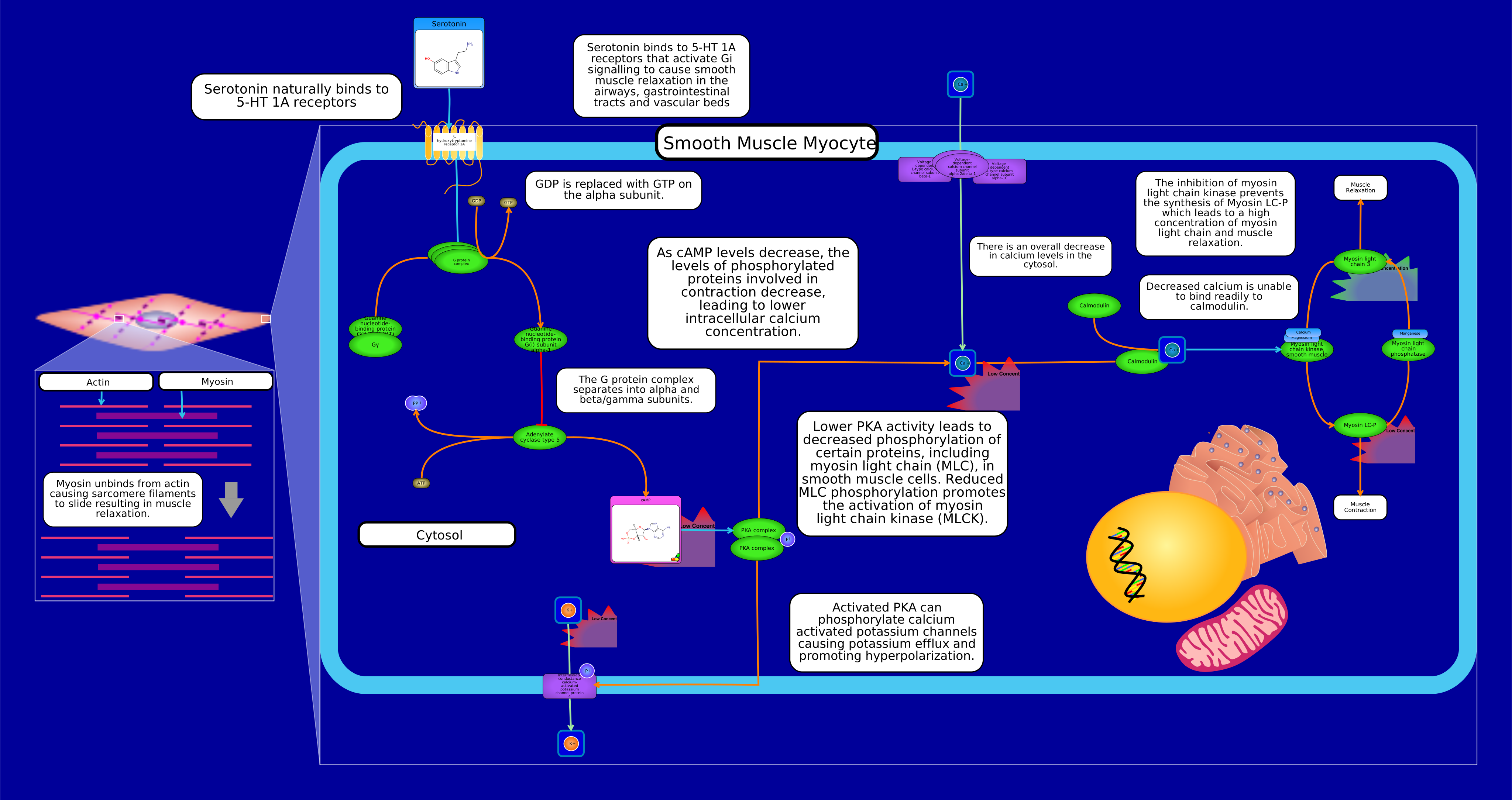 View Pathway
|
physiological
Gi Serotonergic Smooth Muscle RelaxationHomo sapiens
Although numerous GPCRs have the ability to couple to more than one heterotrimeric G protein, a given GPCR is typically classified based on the G protein subfamily (e.g., Gs, Gi/o, or Gq/11) it preferentially activates. Activation of the 5-HT1A receptor typically leads to Gi protein-mediated signaling, which is commonly associated with smooth muscle relaxation rather than contraction. When serotonin (5-HT) or other ligands bind to the 5-HT1A receptor, it triggers a cascade of intracellular events through Gi protein activation. The Gi protein inhibits the activity of adenylate cyclase, which reduces the production of cyclic adenosine monophosphate (cAMP). Reduced cAMP levels can lead to the deactivation of protein kinase A (PKA) and the inhibition of various downstream signaling pathways. In the context of smooth muscle, this reduction in cAMP levels generally leads to smooth muscle relaxation. The physiological effects of serotonin can vary depending on the specific receptors involved, the tissues in question, and the overall context of the signaling pathways. Different serotonin receptors can have opposing effects on smooth muscle, leading to either contraction or relaxation, depending on the receptor subtype and downstream signaling pathways involved. Serotonin increases the motility of the GI tract muscles, induces muscle constriction in the lungs and uterus, influences vessel muscles in both directions (constriction/relaxation), takes part in platelet aggregation, excites nociceptive pain neurons, and influences CNS neurons. Serotonin also plays a role in the symptoms of GI inflammation, acting through different mechanisms to exert pro- or anti-inflammatory activity.
Summarily, Gi inhibits adenylate cyclase 5 which results in the reduced conversion of ATP to cAMP and reduced activation of Protein Kinase A (PKA). Lower PKA activity leads to decreased phosphorylation of certain proteins, including myosin light chain (MLC), in smooth muscle cells. Reduced MLC phosphorylation promotes the activation of myosin light chain kinase (MLCK). Activated PKA can phosphorylate calcium activated potassium channels causing potassium efflux and promoting hyperpolarization. Low potassium levels, and the resulting hyperpolarization, can affect the activity of voltage-gated calcium channels. These channels are involved in calcium influx, which can, in turn, influence cAMP levels and PKA activity. Calcium ions can activate adenylate cyclase, leading to increased cAMP production and PKA activation.
|
Creator: Omolola Created On: September 06, 2023 at 11:12 Last Updated: September 06, 2023 at 11:12 |
PW129305 View Pathway
|
physiological
Gi Serotonergic Smooth Muscle RelaxationMus musculus
Although numerous GPCRs have the ability to couple to more than one heterotrimeric G protein, a given GPCR is typically classified based on the G protein subfamily (e.g., Gs, Gi/o, or Gq/11) it preferentially activates. Activation of the 5-HT1A receptor typically leads to Gi protein-mediated signaling, which is commonly associated with smooth muscle relaxation rather than contraction. When serotonin (5-HT) or other ligands bind to the 5-HT1A receptor, it triggers a cascade of intracellular events through Gi protein activation. The Gi protein inhibits the activity of adenylate cyclase, which reduces the production of cyclic adenosine monophosphate (cAMP). Reduced cAMP levels can lead to the deactivation of protein kinase A (PKA) and the inhibition of various downstream signaling pathways. In the context of smooth muscle, this reduction in cAMP levels generally leads to smooth muscle relaxation. The physiological effects of serotonin can vary depending on the specific receptors involved, the tissues in question, and the overall context of the signaling pathways. Different serotonin receptors can have opposing effects on smooth muscle, leading to either contraction or relaxation, depending on the receptor subtype and downstream signaling pathways involved. Serotonin increases the motility of the GI tract muscles, induces muscle constriction in the lungs and uterus, influences vessel muscles in both directions (constriction/relaxation), takes part in platelet aggregation, excites nociceptive pain neurons, and influences CNS neurons. Serotonin also plays a role in the symptoms of GI inflammation, acting through different mechanisms to exert pro- or anti-inflammatory activity.
Summarily, Gi inhibits adenylate cyclase 5 which results in the reduced conversion of ATP to cAMP and reduced activation of Protein Kinase A (PKA). Lower PKA activity leads to decreased phosphorylation of certain proteins, including myosin light chain (MLC), in smooth muscle cells. Reduced MLC phosphorylation promotes the activation of myosin light chain kinase (MLCK). Activated PKA can phosphorylate calcium activated potassium channels causing potassium efflux and promoting hyperpolarization. Low potassium levels, and the resulting hyperpolarization, can affect the activity of voltage-gated calcium channels. These channels are involved in calcium influx, which can, in turn, influence cAMP levels and PKA activity. Calcium ions can activate adenylate cyclase, leading to increased cAMP production and PKA activation.
|
Creator: Omolola Created On: September 14, 2023 at 10:33 Last Updated: September 14, 2023 at 10:33 |
PW129306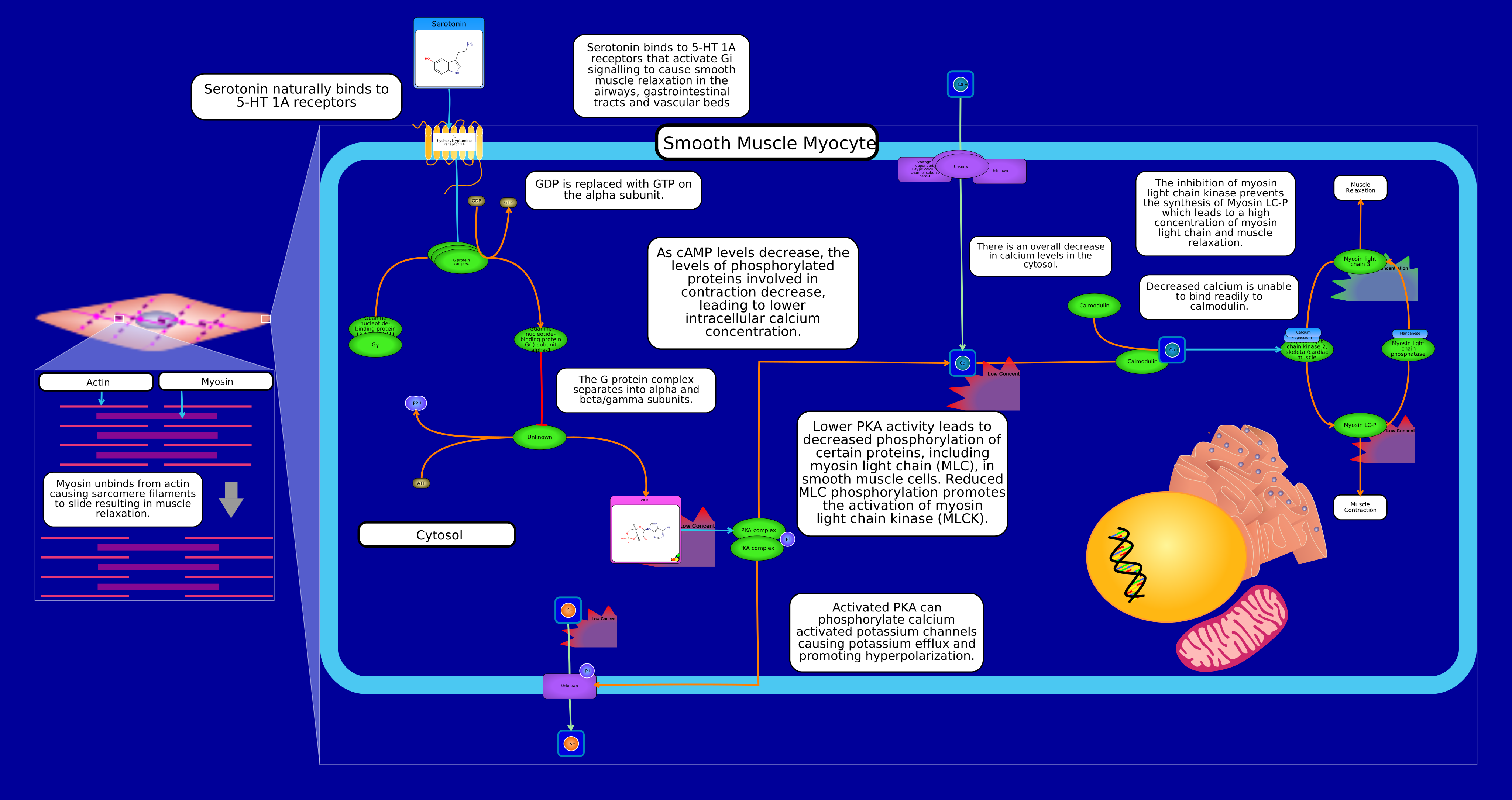 View Pathway
|
physiological
Gi Serotonergic Smooth Muscle RelaxationRattus norvegicus
Although numerous GPCRs have the ability to couple to more than one heterotrimeric G protein, a given GPCR is typically classified based on the G protein subfamily (e.g., Gs, Gi/o, or Gq/11) it preferentially activates. Activation of the 5-HT1A receptor typically leads to Gi protein-mediated signaling, which is commonly associated with smooth muscle relaxation rather than contraction. When serotonin (5-HT) or other ligands bind to the 5-HT1A receptor, it triggers a cascade of intracellular events through Gi protein activation. The Gi protein inhibits the activity of adenylate cyclase, which reduces the production of cyclic adenosine monophosphate (cAMP). Reduced cAMP levels can lead to the deactivation of protein kinase A (PKA) and the inhibition of various downstream signaling pathways. In the context of smooth muscle, this reduction in cAMP levels generally leads to smooth muscle relaxation. The physiological effects of serotonin can vary depending on the specific receptors involved, the tissues in question, and the overall context of the signaling pathways. Different serotonin receptors can have opposing effects on smooth muscle, leading to either contraction or relaxation, depending on the receptor subtype and downstream signaling pathways involved. Serotonin increases the motility of the GI tract muscles, induces muscle constriction in the lungs and uterus, influences vessel muscles in both directions (constriction/relaxation), takes part in platelet aggregation, excites nociceptive pain neurons, and influences CNS neurons. Serotonin also plays a role in the symptoms of GI inflammation, acting through different mechanisms to exert pro- or anti-inflammatory activity.
Summarily, Gi inhibits adenylate cyclase 5 which results in the reduced conversion of ATP to cAMP and reduced activation of Protein Kinase A (PKA). Lower PKA activity leads to decreased phosphorylation of certain proteins, including myosin light chain (MLC), in smooth muscle cells. Reduced MLC phosphorylation promotes the activation of myosin light chain kinase (MLCK). Activated PKA can phosphorylate calcium activated potassium channels causing potassium efflux and promoting hyperpolarization. Low potassium levels, and the resulting hyperpolarization, can affect the activity of voltage-gated calcium channels. These channels are involved in calcium influx, which can, in turn, influence cAMP levels and PKA activity. Calcium ions can activate adenylate cyclase, leading to increased cAMP production and PKA activation.
|
Creator: Omolola Created On: September 14, 2023 at 10:33 Last Updated: September 14, 2023 at 10:33 |
PW128645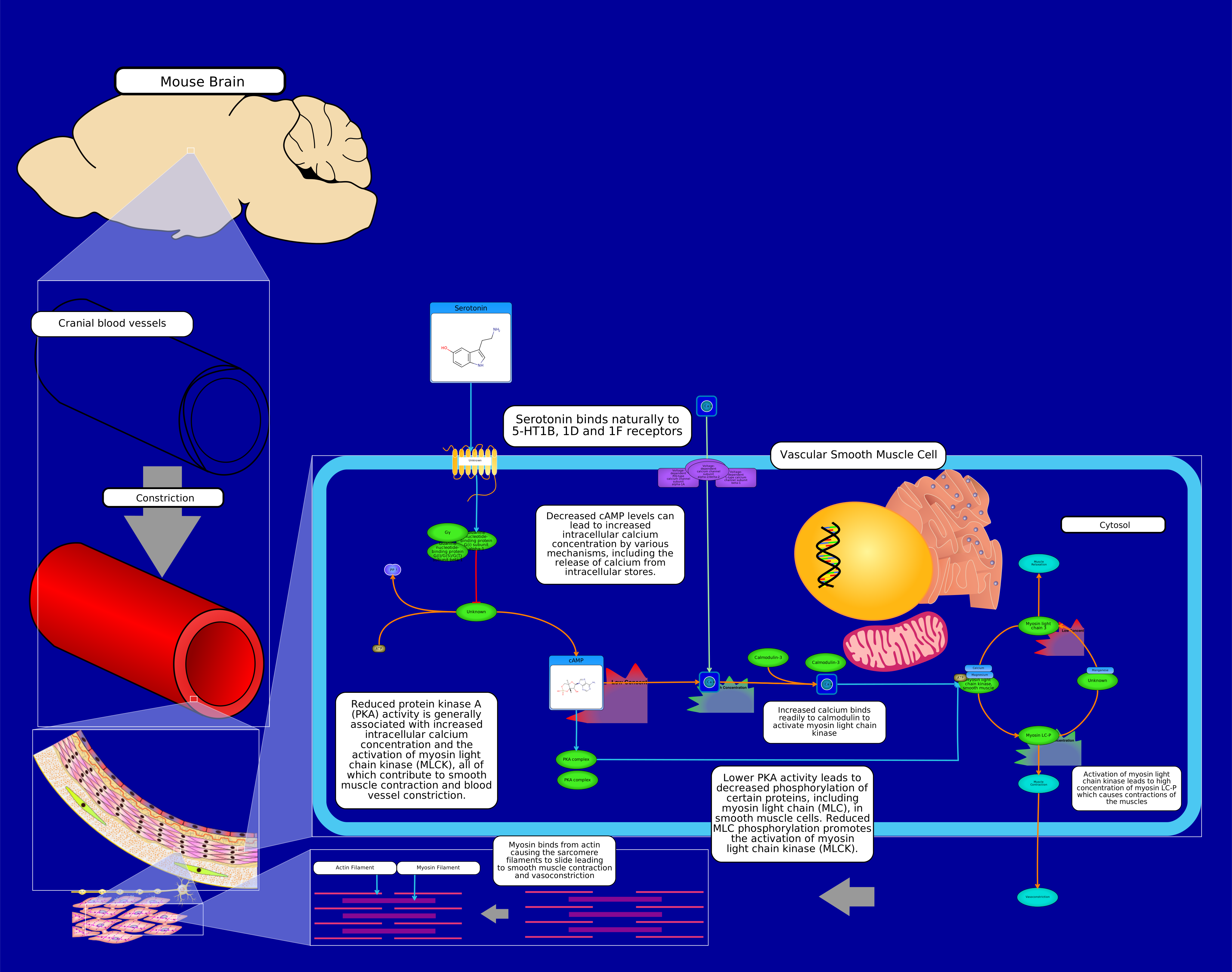 View Pathway
|
physiological
Gi Serotonergic VasoconstrictionMus musculus
G proteins, also known as guanine nucleotide-binding proteins, are a family of proteins that act as molecular switches inside cells, and are involved in transmitting signals from a variety of stimuli outside a cell to its interior. Their activity is regulated by factors that control their ability to bind to and hydrolyze guanosine triphosphate (GTP) to guanosine diphosphate (GDP). When they are bound to GTP, they are 'on', and, when they are bound to GDP, they are 'off'. G proteins belong to the larger group of enzymes called GTPases. Heterotrimeric G proteins located within the cell are activated by G protein-coupled receptors (GPCRs) that span the cell membrane. Signaling molecules bind to a domain of the GPCR located outside the cell, and an intracellular GPCR domain then in turn activates a particular G protein. Some active-state GPCRs have also been shown to be "pre-coupled" with G proteins, whereas in other cases a collision coupling mechanism is thought to occur. The G protein triggers a cascade of further signaling events that finally results in a change in cell function. G protein-coupled receptors and G proteins working together transmit signals from many hormones, neurotransmitters, and other signaling factors. G proteins regulate metabolic enzymes, ion channels, transporter proteins, and other parts of the cell machinery, controlling transcription, motility, contractility, and secretion, which in turn regulate diverse systemic functions such as embryonic development, learning and memory, and homeostasis. Receptor-activated G proteins are bound to the inner surface of the cell membrane. They consist of the Gα and the tightly associated Gβγ subunits. There are four main families of Gα subunits: Gαs (G stimulatory), Gαi (G inhibitory), Gαq/11, and Gα12/13. They behave differently in the recognition of the effector molecule, but share a similar mechanism of activation. When a ligand activates the G protein-coupled receptor, it induces a conformational change in the receptor that allows the receptor to function as a guanine nucleotide exchange factor (GEF) that exchanges GDP for GTP. The GTP (or GDP) is bound to the Gα subunit in the traditional view of heterotrimeric GPCR activation. This exchange triggers the dissociation of the Gα subunit (which is bound to GTP) from the Gβγ dimer and the receptor as a whole. Both Gα-GTP and Gβγ can then activate different signaling cascades (or second messenger pathways) and effector proteins, while the receptor is able to activate the next G protein. Gi protein alpha subunit is a family of heterotrimeric G protein alpha subunits. This family is also commonly called the Gi/o (Gi /Go ) family or Gi/o/z/t family to include closely related family members. G alpha subunits may be referred to as Gi alpha, Gαi, or Giα. Gi proteins primarily inhibit the cAMP dependent pathway by inhibiting adenylyl cyclase activity, decreasing the production of cAMP from ATP, which, in turn, results in decreased activity of cAMP-dependent protein kinase. Therefore, the ultimate effect of Gi is the inhibition of the cAMP-dependent protein kinase. The Gβγ liberated by activation of Gi and Go proteins is particularly able to activate downstream signaling to effectors such as G protein-coupled inwardly-rectifying potassium channels (GIRKs). Gi and Go proteins are substrates for pertussis toxin, produced by Bordetella pertussis, the infectious agent in whooping cough. Pertussis toxin is an ADP-ribosylase enzyme that adds an ADP-ribose moiety to a particular cysteine residue in Giα and Goα proteins, preventing their coupling to and activation by GPCRs, thus turning off Gi and Go cell signaling pathways. Activation of Gi proteins in vascular smooth muscle cells often results in vasoconstriction. This is because reduced cAMP levels and decreased PKA activity lead to increased intracellular calcium concentrations, promoting smooth muscle contraction. In summary, while Gi signaling plays a role in both smooth and cardiac muscle tissues, its specific effects and functions differ due to the distinct roles and regulatory mechanisms of these muscles. In smooth muscle, Gi signaling often leads to vasoconstriction, while in cardiac muscle, it primarily regulates heart rate through the inhibition of If channels in the SA node, resulting in a negative chronotropic effect.
Summarily, Vasoconstriction and muscle contraction occurs due to the inhibition of adenylate cyclase. Gi inhibits adenylate cyclase 5 which results in the reduced conversion of ATP to cAMP and reduced activation of Protein Kinase A (PKA). Lower PKA activity leads to decreased phosphorylation of certain proteins, including myosin light chain (MLC), in smooth muscle cells. Reduced MLC phosphorylation promotes the activation of myosin light chain kinase (MLCK). Activated PKA can phosphorylate calcium activated potassium channels causing potassium efflux and promoting hyperpolarization. Low potassium levels, and the resulting hyperpolarization, can affect the activity of voltage-gated calcium channels. These channels are involved in calcium influx, which can, in turn, influence cAMP levels and PKA activity. Calcium ions can activate adenylate cyclase, leading to increased cAMP production and PKA activation.
|
Creator: Omolola Created On: September 08, 2023 at 09:38 Last Updated: September 08, 2023 at 09:38 |
PW128610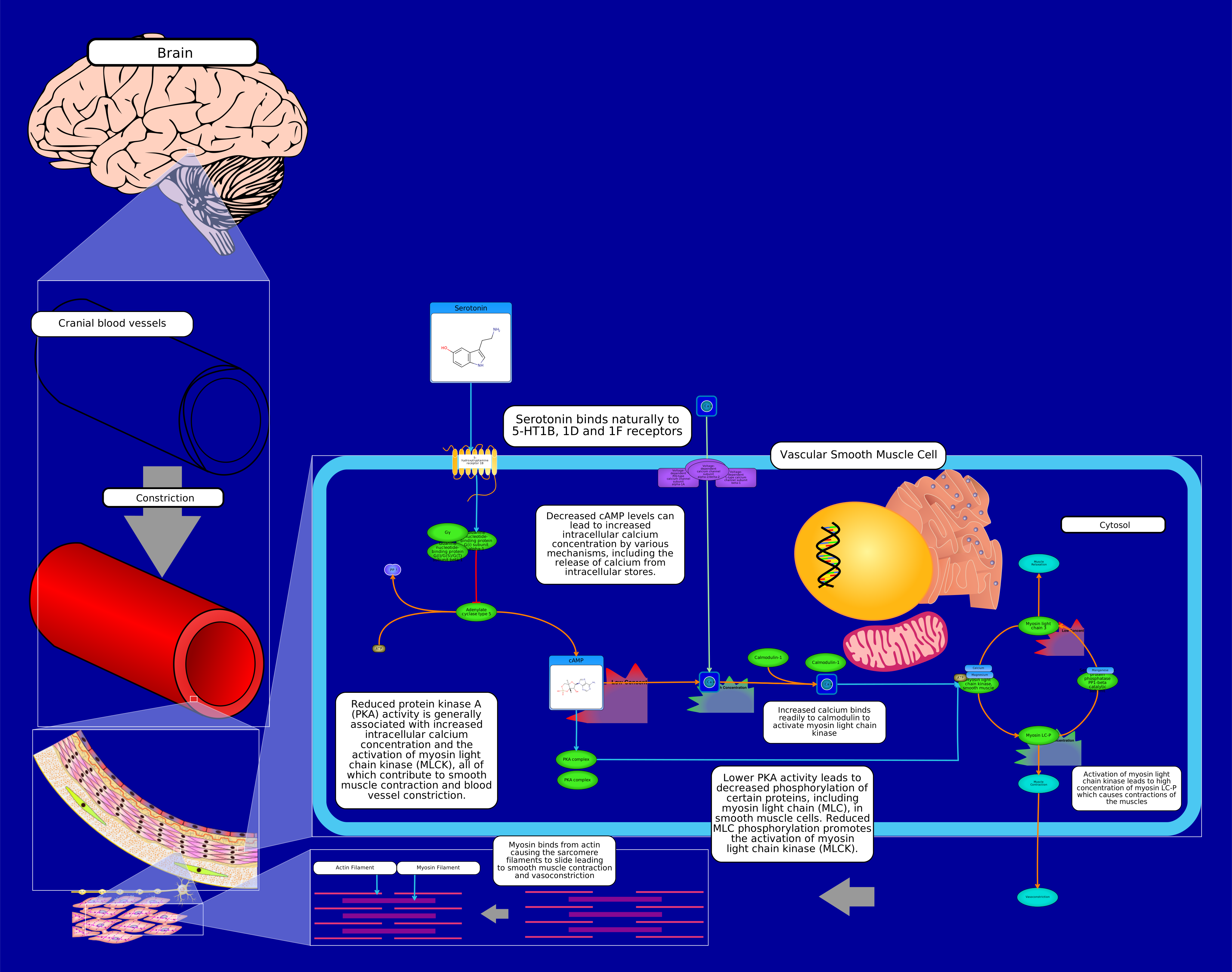 View Pathway
|
drug action
Gi Serotonergic VasoconstrictionHomo sapiens
G proteins, also known as guanine nucleotide-binding proteins, are a family of proteins that act as molecular switches inside cells, and are involved in transmitting signals from a variety of stimuli outside a cell to its interior. Their activity is regulated by factors that control their ability to bind to and hydrolyze guanosine triphosphate (GTP) to guanosine diphosphate (GDP). When they are bound to GTP, they are 'on', and, when they are bound to GDP, they are 'off'. G proteins belong to the larger group of enzymes called GTPases. Heterotrimeric G proteins located within the cell are activated by G protein-coupled receptors (GPCRs) that span the cell membrane. Signaling molecules bind to a domain of the GPCR located outside the cell, and an intracellular GPCR domain then in turn activates a particular G protein. Some active-state GPCRs have also been shown to be "pre-coupled" with G proteins, whereas in other cases a collision coupling mechanism is thought to occur. The G protein triggers a cascade of further signaling events that finally results in a change in cell function. G protein-coupled receptors and G proteins working together transmit signals from many hormones, neurotransmitters, and other signaling factors. G proteins regulate metabolic enzymes, ion channels, transporter proteins, and other parts of the cell machinery, controlling transcription, motility, contractility, and secretion, which in turn regulate diverse systemic functions such as embryonic development, learning and memory, and homeostasis. Receptor-activated G proteins are bound to the inner surface of the cell membrane. They consist of the Gα and the tightly associated Gβγ subunits. There are four main families of Gα subunits: Gαs (G stimulatory), Gαi (G inhibitory), Gαq/11, and Gα12/13. They behave differently in the recognition of the effector molecule, but share a similar mechanism of activation. When a ligand activates the G protein-coupled receptor, it induces a conformational change in the receptor that allows the receptor to function as a guanine nucleotide exchange factor (GEF) that exchanges GDP for GTP. The GTP (or GDP) is bound to the Gα subunit in the traditional view of heterotrimeric GPCR activation. This exchange triggers the dissociation of the Gα subunit (which is bound to GTP) from the Gβγ dimer and the receptor as a whole. Both Gα-GTP and Gβγ can then activate different signaling cascades (or second messenger pathways) and effector proteins, while the receptor is able to activate the next G protein. Gi protein alpha subunit is a family of heterotrimeric G protein alpha subunits. This family is also commonly called the Gi/o (Gi /Go ) family or Gi/o/z/t family to include closely related family members. G alpha subunits may be referred to as Gi alpha, Gαi, or Giα. Gi proteins primarily inhibit the cAMP dependent pathway by inhibiting adenylyl cyclase activity, decreasing the production of cAMP from ATP, which, in turn, results in decreased activity of cAMP-dependent protein kinase. Therefore, the ultimate effect of Gi is the inhibition of the cAMP-dependent protein kinase. The Gβγ liberated by activation of Gi and Go proteins is particularly able to activate downstream signaling to effectors such as G protein-coupled inwardly-rectifying potassium channels (GIRKs). Gi and Go proteins are substrates for pertussis toxin, produced by Bordetella pertussis, the infectious agent in whooping cough. Pertussis toxin is an ADP-ribosylase enzyme that adds an ADP-ribose moiety to a particular cysteine residue in Giα and Goα proteins, preventing their coupling to and activation by GPCRs, thus turning off Gi and Go cell signaling pathways. Activation of Gi proteins in vascular smooth muscle cells often results in vasoconstriction. This is because reduced cAMP levels and decreased PKA activity lead to increased intracellular calcium concentrations, promoting smooth muscle contraction. In summary, while Gi signaling plays a role in both smooth and cardiac muscle tissues, its specific effects and functions differ due to the distinct roles and regulatory mechanisms of these muscles. In smooth muscle, Gi signaling often leads to vasoconstriction, while in cardiac muscle, it primarily regulates heart rate through the inhibition of If channels in the SA node, resulting in a negative chronotropic effect.
Summarily, Vasoconstriction and muscle contraction occurs due to the inhibition of adenylate cyclase. Gi inhibits adenylate cyclase 5 which results in the reduced conversion of ATP to cAMP and reduced activation of Protein Kinase A (PKA). Lower PKA activity leads to decreased phosphorylation of certain proteins, including myosin light chain (MLC), in smooth muscle cells. Reduced MLC phosphorylation promotes the activation of myosin light chain kinase (MLCK). Activated PKA can phosphorylate calcium activated potassium channels causing potassium efflux and promoting hyperpolarization. Low potassium levels, and the resulting hyperpolarization, can affect the activity of voltage-gated calcium channels. These channels are involved in calcium influx, which can, in turn, influence cAMP levels and PKA activity. Calcium ions can activate adenylate cyclase, leading to increased cAMP production and PKA activation.
|
Creator: Omolola Created On: September 06, 2023 at 15:36 Last Updated: September 06, 2023 at 15:36 |
PW128646 View Pathway
|
physiological
Gi Serotonergic VasoconstrictionBos taurus
Serotonin has the potential to serve as an essential tool in the prevention of transition-related dairy cow diseases. Serotonin is a potent regulator of both calcium homeostasis and energy homeostasis during lactation in both rodent and dairy cow models. G proteins, also known as guanine nucleotide-binding proteins, are a family of proteins that act as molecular switches inside cells, and are involved in transmitting signals from a variety of stimuli outside a cell to its interior. Their activity is regulated by factors that control their ability to bind to and hydrolyze guanosine triphosphate (GTP) to guanosine diphosphate (GDP). When they are bound to GTP, they are 'on', and, when they are bound to GDP, they are 'off'. G proteins belong to the larger group of enzymes called GTPases. Heterotrimeric G proteins located within the cell are activated by G protein-coupled receptors (GPCRs) that span the cell membrane. Signaling molecules bind to a domain of the GPCR located outside the cell, and an intracellular GPCR domain then in turn activates a particular G protein. Some active-state GPCRs have also been shown to be "pre-coupled" with G proteins, whereas in other cases a collision coupling mechanism is thought to occur. The G protein triggers a cascade of further signaling events that finally results in a change in cell function. G protein-coupled receptors and G proteins working together transmit signals from many hormones, neurotransmitters, and other signaling factors. G proteins regulate metabolic enzymes, ion channels, transporter proteins, and other parts of the cell machinery, controlling transcription, motility, contractility, and secretion, which in turn regulate diverse systemic functions such as embryonic development, learning and memory, and homeostasis. Receptor-activated G proteins are bound to the inner surface of the cell membrane. They consist of the Gα and the tightly associated Gβγ subunits. There are four main families of Gα subunits: Gαs (G stimulatory), Gαi (G inhibitory), Gαq/11, and Gα12/13. They behave differently in the recognition of the effector molecule, but share a similar mechanism of activation. When a ligand activates the G protein-coupled receptor, it induces a conformational change in the receptor that allows the receptor to function as a guanine nucleotide exchange factor (GEF) that exchanges GDP for GTP. The GTP (or GDP) is bound to the Gα subunit in the traditional view of heterotrimeric GPCR activation. This exchange triggers the dissociation of the Gα subunit (which is bound to GTP) from the Gβγ dimer and the receptor as a whole. Both Gα-GTP and Gβγ can then activate different signaling cascades (or second messenger pathways) and effector proteins, while the receptor is able to activate the next G protein. Gi protein alpha subunit is a family of heterotrimeric G protein alpha subunits. This family is also commonly called the Gi/o (Gi /Go ) family or Gi/o/z/t family to include closely related family members. G alpha subunits may be referred to as Gi alpha, Gαi, or Giα. Gi proteins primarily inhibit the cAMP dependent pathway by inhibiting adenylyl cyclase activity, decreasing the production of cAMP from ATP, which, in turn, results in decreased activity of cAMP-dependent protein kinase. Therefore, the ultimate effect of Gi is the inhibition of the cAMP-dependent protein kinase. The Gβγ liberated by activation of Gi and Go proteins is particularly able to activate downstream signaling to effectors such as G protein-coupled inwardly-rectifying potassium channels (GIRKs). Gi and Go proteins are substrates for pertussis toxin, produced by Bordetella pertussis, the infectious agent in whooping cough. Pertussis toxin is an ADP-ribosylase enzyme that adds an ADP-ribose moiety to a particular cysteine residue in Giα and Goα proteins, preventing their coupling to and activation by GPCRs, thus turning off Gi and Go cell signaling pathways. Activation of Gi proteins in vascular smooth muscle cells often results in vasoconstriction. This is because reduced cAMP levels and decreased PKA activity lead to increased intracellular calcium concentrations, promoting smooth muscle contraction. In summary, while Gi signaling plays a role in both smooth and cardiac muscle tissues, its specific effects and functions differ due to the distinct roles and regulatory mechanisms of these muscles. In smooth muscle, Gi signaling often leads to vasoconstriction, while in cardiac muscle, it primarily regulates heart rate through the inhibition of If channels in the SA node, resulting in a negative chronotropic effect.
Summarily, Vasoconstriction and muscle contraction occurs due to the inhibition of adenylate cyclase. Gi inhibits adenylate cyclase 5 which results in the reduced conversion of ATP to cAMP and reduced activation of Protein Kinase A (PKA). Lower PKA activity leads to decreased phosphorylation of certain proteins, including myosin light chain (MLC), in smooth muscle cells. Reduced MLC phosphorylation promotes the activation of myosin light chain kinase (MLCK). Activated PKA can phosphorylate calcium activated potassium channels causing potassium efflux and promoting hyperpolarization. Low potassium levels, and the resulting hyperpolarization, can affect the activity of voltage-gated calcium channels. These channels are involved in calcium influx, which can, in turn, influence cAMP levels and PKA activity. Calcium ions can activate adenylate cyclase, leading to increased cAMP production and PKA activation.
|
Creator: Omolola Created On: September 08, 2023 at 10:19 Last Updated: September 08, 2023 at 10:19 |
PW128644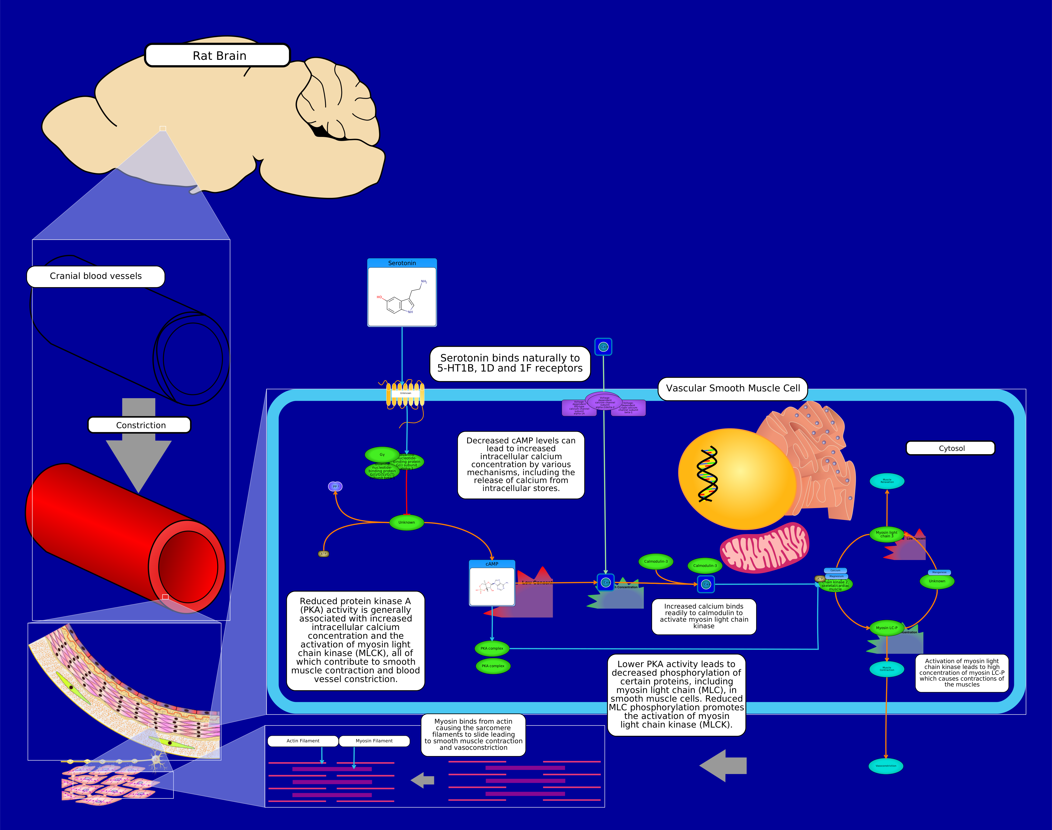 View Pathway
|
physiological
Gi Serotonergic VasoconstrictionRattus norvegicus
G proteins, also known as guanine nucleotide-binding proteins, are a family of proteins that act as molecular switches inside cells, and are involved in transmitting signals from a variety of stimuli outside a cell to its interior. Their activity is regulated by factors that control their ability to bind to and hydrolyze guanosine triphosphate (GTP) to guanosine diphosphate (GDP). When they are bound to GTP, they are 'on', and, when they are bound to GDP, they are 'off'. G proteins belong to the larger group of enzymes called GTPases. Heterotrimeric G proteins located within the cell are activated by G protein-coupled receptors (GPCRs) that span the cell membrane. Signaling molecules bind to a domain of the GPCR located outside the cell, and an intracellular GPCR domain then in turn activates a particular G protein. Some active-state GPCRs have also been shown to be "pre-coupled" with G proteins, whereas in other cases a collision coupling mechanism is thought to occur. The G protein triggers a cascade of further signaling events that finally results in a change in cell function. G protein-coupled receptors and G proteins working together transmit signals from many hormones, neurotransmitters, and other signaling factors. G proteins regulate metabolic enzymes, ion channels, transporter proteins, and other parts of the cell machinery, controlling transcription, motility, contractility, and secretion, which in turn regulate diverse systemic functions such as embryonic development, learning and memory, and homeostasis. Receptor-activated G proteins are bound to the inner surface of the cell membrane. They consist of the Gα and the tightly associated Gβγ subunits. There are four main families of Gα subunits: Gαs (G stimulatory), Gαi (G inhibitory), Gαq/11, and Gα12/13. They behave differently in the recognition of the effector molecule, but share a similar mechanism of activation. When a ligand activates the G protein-coupled receptor, it induces a conformational change in the receptor that allows the receptor to function as a guanine nucleotide exchange factor (GEF) that exchanges GDP for GTP. The GTP (or GDP) is bound to the Gα subunit in the traditional view of heterotrimeric GPCR activation. This exchange triggers the dissociation of the Gα subunit (which is bound to GTP) from the Gβγ dimer and the receptor as a whole. Both Gα-GTP and Gβγ can then activate different signaling cascades (or second messenger pathways) and effector proteins, while the receptor is able to activate the next G protein. Gi protein alpha subunit is a family of heterotrimeric G protein alpha subunits. This family is also commonly called the Gi/o (Gi /Go ) family or Gi/o/z/t family to include closely related family members. G alpha subunits may be referred to as Gi alpha, Gαi, or Giα. Gi proteins primarily inhibit the cAMP dependent pathway by inhibiting adenylyl cyclase activity, decreasing the production of cAMP from ATP, which, in turn, results in decreased activity of cAMP-dependent protein kinase. Therefore, the ultimate effect of Gi is the inhibition of the cAMP-dependent protein kinase. The Gβγ liberated by activation of Gi and Go proteins is particularly able to activate downstream signaling to effectors such as G protein-coupled inwardly-rectifying potassium channels (GIRKs). Gi and Go proteins are substrates for pertussis toxin, produced by Bordetella pertussis, the infectious agent in whooping cough. Pertussis toxin is an ADP-ribosylase enzyme that adds an ADP-ribose moiety to a particular cysteine residue in Giα and Goα proteins, preventing their coupling to and activation by GPCRs, thus turning off Gi and Go cell signaling pathways. Activation of Gi proteins in vascular smooth muscle cells often results in vasoconstriction. This is because reduced cAMP levels and decreased PKA activity lead to increased intracellular calcium concentrations, promoting smooth muscle contraction. In summary, while Gi signaling plays a role in both smooth and cardiac muscle tissues, its specific effects and functions differ due to the distinct roles and regulatory mechanisms of these muscles. In smooth muscle, Gi signaling often leads to vasoconstriction, while in cardiac muscle, it primarily regulates heart rate through the inhibition of If channels in the SA node, resulting in a negative chronotropic effect.
Summarily, Vasoconstriction and muscle contraction occurs due to the inhibition of adenylate cyclase. Gi inhibits adenylate cyclase 5 which results in the reduced conversion of ATP to cAMP and reduced activation of Protein Kinase A (PKA). Lower PKA activity leads to decreased phosphorylation of certain proteins, including myosin light chain (MLC), in smooth muscle cells. Reduced MLC phosphorylation promotes the activation of myosin light chain kinase (MLCK). Activated PKA can phosphorylate calcium activated potassium channels causing potassium efflux and promoting hyperpolarization. Low potassium levels, and the resulting hyperpolarization, can affect the activity of voltage-gated calcium channels. These channels are involved in calcium influx, which can, in turn, influence cAMP levels and PKA activity. Calcium ions can activate adenylate cyclase, leading to increased cAMP production and PKA activation.
|
Creator: Omolola Created On: September 08, 2023 at 09:38 Last Updated: September 08, 2023 at 09:38 |
PW012930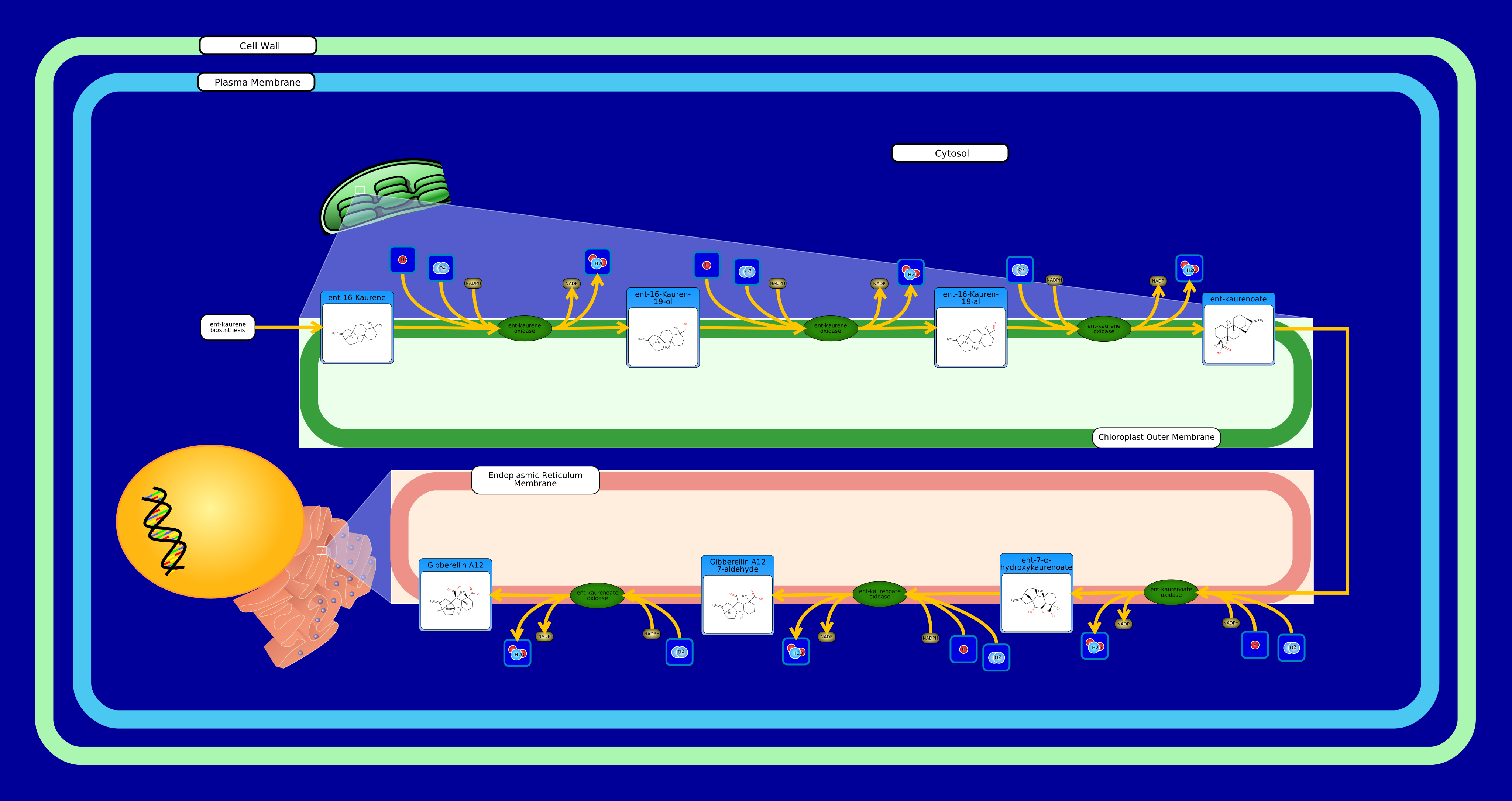 View Pathway
|
Gibberellin A12 BiosynthesisArabidopsis thaliana
Gibberellins (GAs) are a large class of tetracyclic diterpenoid plant hormones that regulate numerous growth and developmental processes, such as seed germination, organ elongation, and flowering induction. All known gibberellins share an ent-gibberellane skeleton and follow the same synthesis pathway. Biosynthesis begins in the plasmids via the terpenoid pathway and finishes in the endoplasmic reticulum and cytosol where they undergo modification until a biologically-active form is reached (GA1, GA3, GA4, or GA7). Gibberellins are named in the order that they are discovered (GA1 through GAn). Serving as a branch point, the first true gibberellin GA12 is used to synthesize the full range of gibberellins by undergoing a multitude of oxidations and cyclizations. Gibberellin A12 biosynthesis, beginning at the chloroplast outer membrane and finishing at the endoplasmic reticulum membrane, comprises of six oxidation steps catalyzed by two membrane-associated multifunctional enzymes of the cytochrome P450 family: ent-kaurene oxidase and ent-kaurenoic acid oxidase. ent-Kaurene oxidase converts ent-kaurene into ent-kaurenoate via three successive oxidations of the 4-methyl group. ent-Kaurenoate oxidase converts ent-kaurenoate into gibberellin A12 via three successive oxidations at carbon positions C-7 and C-6 in gibberellin A12 biosynthesis.
|
Creator: Carin Li Created On: March 02, 2017 at 00:46 Last Updated: March 02, 2017 at 00:46 |
PW123683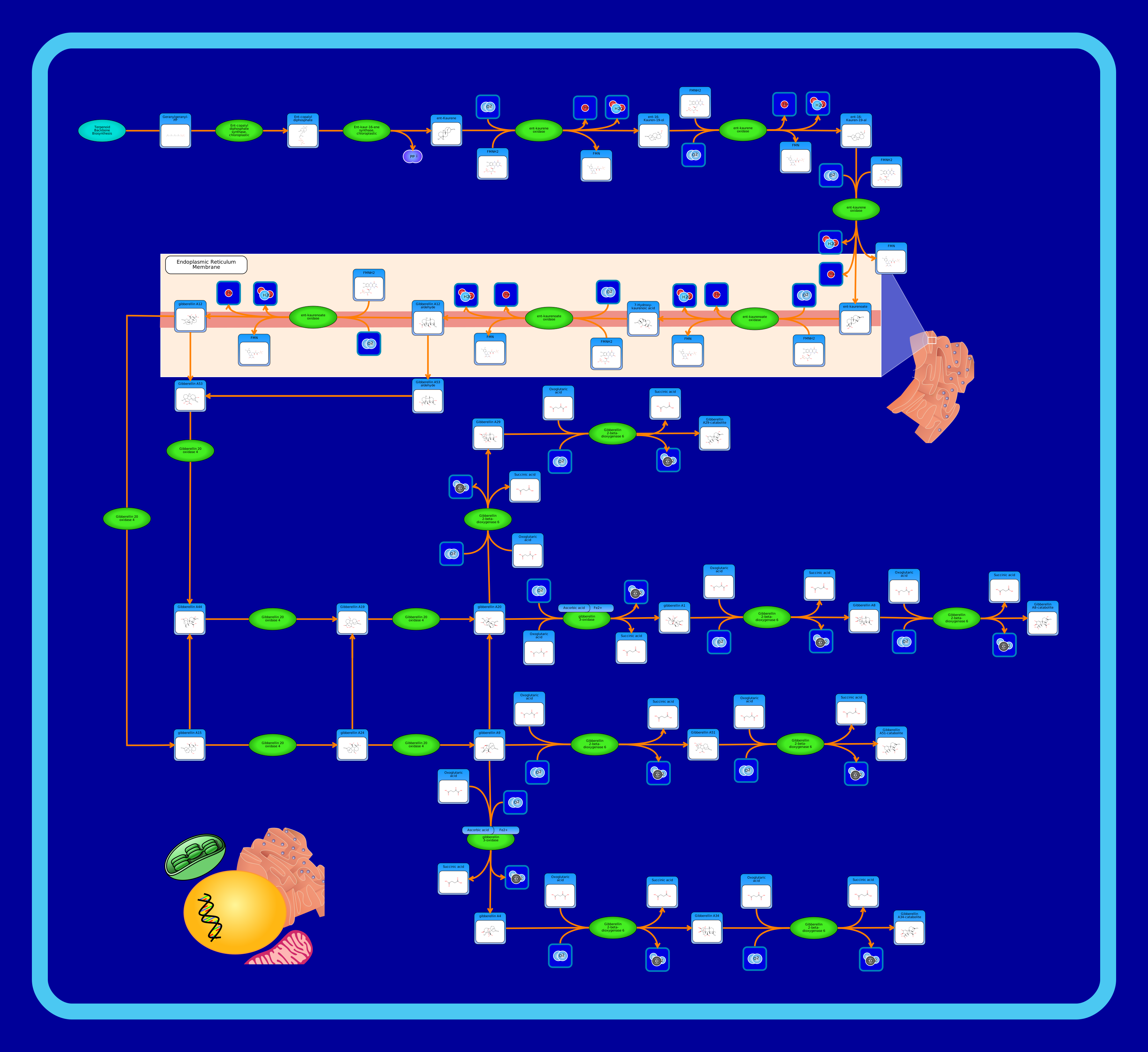 View Pathway
|
Gibberellin and Gibberellin Precursor Biosynthesis PathwayArabidopsis thaliana
A gibberellin is a plant hormone that is necessary for many functions within a plant. Some of these functions include shoot length, cell division, and bolting induction. This pathway shows the biosynthesis of gibberellin precursors, within the larger diterpenoid biosynthesis pathway. It begins with geranygeranyl-PP, synthesized from Terpenoid backbone biosynthesis, using the enzyme ent-copalyl diphosphate synthase to create ent-copalyl diphosphate. From here, ent-copalyl diphosphate uses ent-kaur-16-ene synthase to create ent-kaurene. Ent-kaurene continues this pathway, teaming up with ent-kaurene oxidase to produce ent-16-kauren-19-ol. Ent-kaurene oxidase is used again in combination with ent-16-kauren-19-ol to synthesize ent-16-kauren-19-al. The enzyme ent-kaurene oxidase is used once more, to create ent-kaurenoate. At this point in the pathway, the reactions begin to take place in the endoplasmic reticulum membrane. Here, ent-kaurenoae oxidase converts ent-kaurenoate into 7-hydroxy-kaurenoic acid. This acid then uses ent-kaurenoate oxidase again to become gibberellin A12 aldehyde. Exiting the endoplasmic reticulum membrane, gibberellin A12 uses gibberellin 20 oxidase 4 to produce gibberellin A15. The reactions continue, still using gibberellin 20 oxidase 4 to create gibberellin A24, and subsequently gibberellin A9. After this, gibberellin a9 is converted to gibberellin a51 through the enzyme gibberellin 2-beta-dioxygenase 6. Gibberellin A51 then uses this same enzyme to create a gibberellin A51-catabolite. There are many offshoots in this pathway that are not described in detail, but all assume a similar chain of reactions and ultimately result in the production of different gibberellin catabolites.
|
Creator: Maïlys Paccoud Created On: January 11, 2020 at 14:41 Last Updated: January 11, 2020 at 14:41 |