
Browsing Pathways
Showing 541201 -
541210 of 605359 pathways
| PathBank ID | Pathway Name and Description | Pathway Class | Chemical Compounds | Proteins |
|---|---|---|---|---|
SMP0131619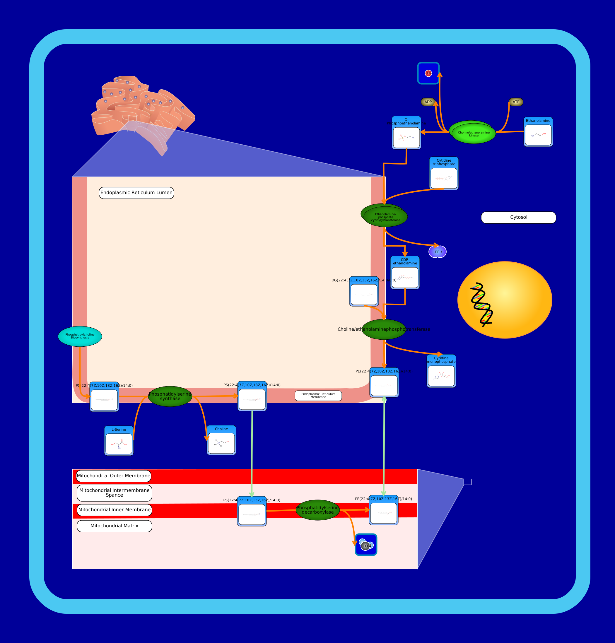 View Pathway
|
Phosphatidylethanolamine Biosynthesis PE(22:4(7Z,10Z,13Z,16Z)/14:0)Mus musculus
Phosphatidylethanolamines (PE) are a class of phospholipids that incorporate a phosphoric acid headgroup into a diacylglycerol backbone. They are the second most abundant phospholipid in eukaryotic cell membranes, and contrary to phosphatidylcholine, it is concentrated with phosphatidylserine in the cell membrane's inner leaflet. In Homo sapiens, there exist two phosphatidylethanolamine biosynthesis pathways. In the visualization, all enzymes that are dark green in colour are membrane-localized. The first pathway synthesizes phosphatidylethanolamine from ethanolamine via the Kennedy pathway. First, the cytosol-localized enzyme choline/ethanolamine kinase catalyzes the conversion of choline into phosphocholine. Second, choline-phosphate cytidylyltransferase, localized to the endoplasmic reticulum membrane, catalyzes the conversion of phosphocholine to CDP-choline. Last, choline/ethanolaminephosphotransferase catalyzes phosphatidylcholine biosynthesis from CDP-choline. It requires either magnesium or manganese ions as cofactors. Phosphatidylethanolamine is also synthesized from phosphatidylserine at the mitochondrial inner membrane by phosphatidylserine decarboxylase. Phosphatidylserine, itself, is synthesized using a base-exchange reaction with phosphatidylcholine. This reaction is catalyzed by phosphatidylserine synthase which is located in the endoplasmic reticulum membrane.
|
Metabolite
Metabolic
|
|
|
SMP0131602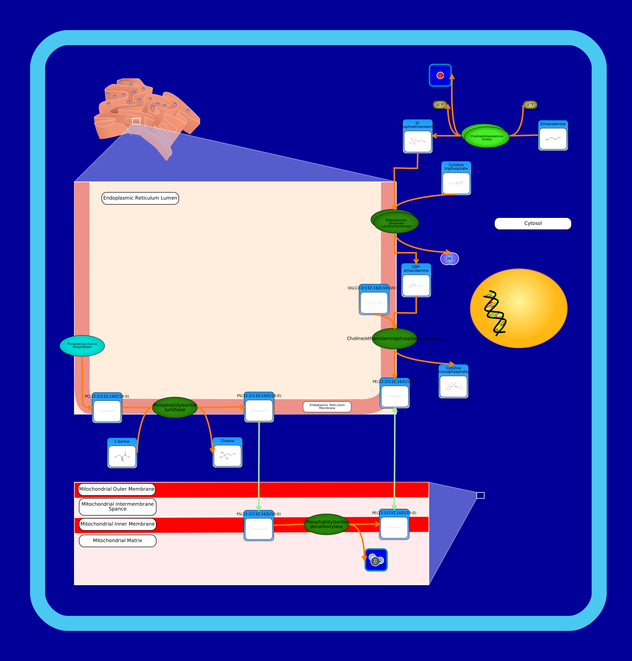 View Pathway
|
Phosphatidylethanolamine Biosynthesis PE(22:2(13Z,16Z)/18:0)Mus musculus
Phosphatidylethanolamines (PE) are a class of phospholipids that incorporate a phosphoric acid headgroup into a diacylglycerol backbone. They are the second most abundant phospholipid in eukaryotic cell membranes, and contrary to phosphatidylcholine, it is concentrated with phosphatidylserine in the cell membrane's inner leaflet. In Homo sapiens, there exist two phosphatidylethanolamine biosynthesis pathways. In the visualization, all enzymes that are dark green in colour are membrane-localized. The first pathway synthesizes phosphatidylethanolamine from ethanolamine via the Kennedy pathway. First, the cytosol-localized enzyme choline/ethanolamine kinase catalyzes the conversion of choline into phosphocholine. Second, choline-phosphate cytidylyltransferase, localized to the endoplasmic reticulum membrane, catalyzes the conversion of phosphocholine to CDP-choline. Last, choline/ethanolaminephosphotransferase catalyzes phosphatidylcholine biosynthesis from CDP-choline. It requires either magnesium or manganese ions as cofactors. Phosphatidylethanolamine is also synthesized from phosphatidylserine at the mitochondrial inner membrane by phosphatidylserine decarboxylase. Phosphatidylserine, itself, is synthesized using a base-exchange reaction with phosphatidylcholine. This reaction is catalyzed by phosphatidylserine synthase which is located in the endoplasmic reticulum membrane.
|
Metabolite
Metabolic
|
|
|
SMP0131583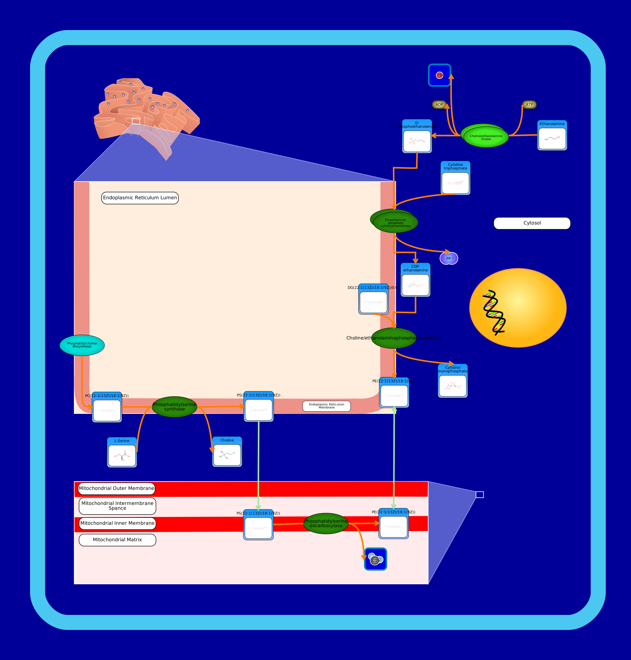 View Pathway
|
Phosphatidylethanolamine Biosynthesis PE(22:1(13Z)/18:1(9Z))Mus musculus
Phosphatidylethanolamines (PE) are a class of phospholipids that incorporate a phosphoric acid headgroup into a diacylglycerol backbone. They are the second most abundant phospholipid in eukaryotic cell membranes, and contrary to phosphatidylcholine, it is concentrated with phosphatidylserine in the cell membrane's inner leaflet. In Homo sapiens, there exist two phosphatidylethanolamine biosynthesis pathways. In the visualization, all enzymes that are dark green in colour are membrane-localized. The first pathway synthesizes phosphatidylethanolamine from ethanolamine via the Kennedy pathway. First, the cytosol-localized enzyme choline/ethanolamine kinase catalyzes the conversion of choline into phosphocholine. Second, choline-phosphate cytidylyltransferase, localized to the endoplasmic reticulum membrane, catalyzes the conversion of phosphocholine to CDP-choline. Last, choline/ethanolaminephosphotransferase catalyzes phosphatidylcholine biosynthesis from CDP-choline. It requires either magnesium or manganese ions as cofactors. Phosphatidylethanolamine is also synthesized from phosphatidylserine at the mitochondrial inner membrane by phosphatidylserine decarboxylase. Phosphatidylserine, itself, is synthesized using a base-exchange reaction with phosphatidylcholine. This reaction is catalyzed by phosphatidylserine synthase which is located in the endoplasmic reticulum membrane.
|
Metabolite
Metabolic
|
|
|
SMP0131588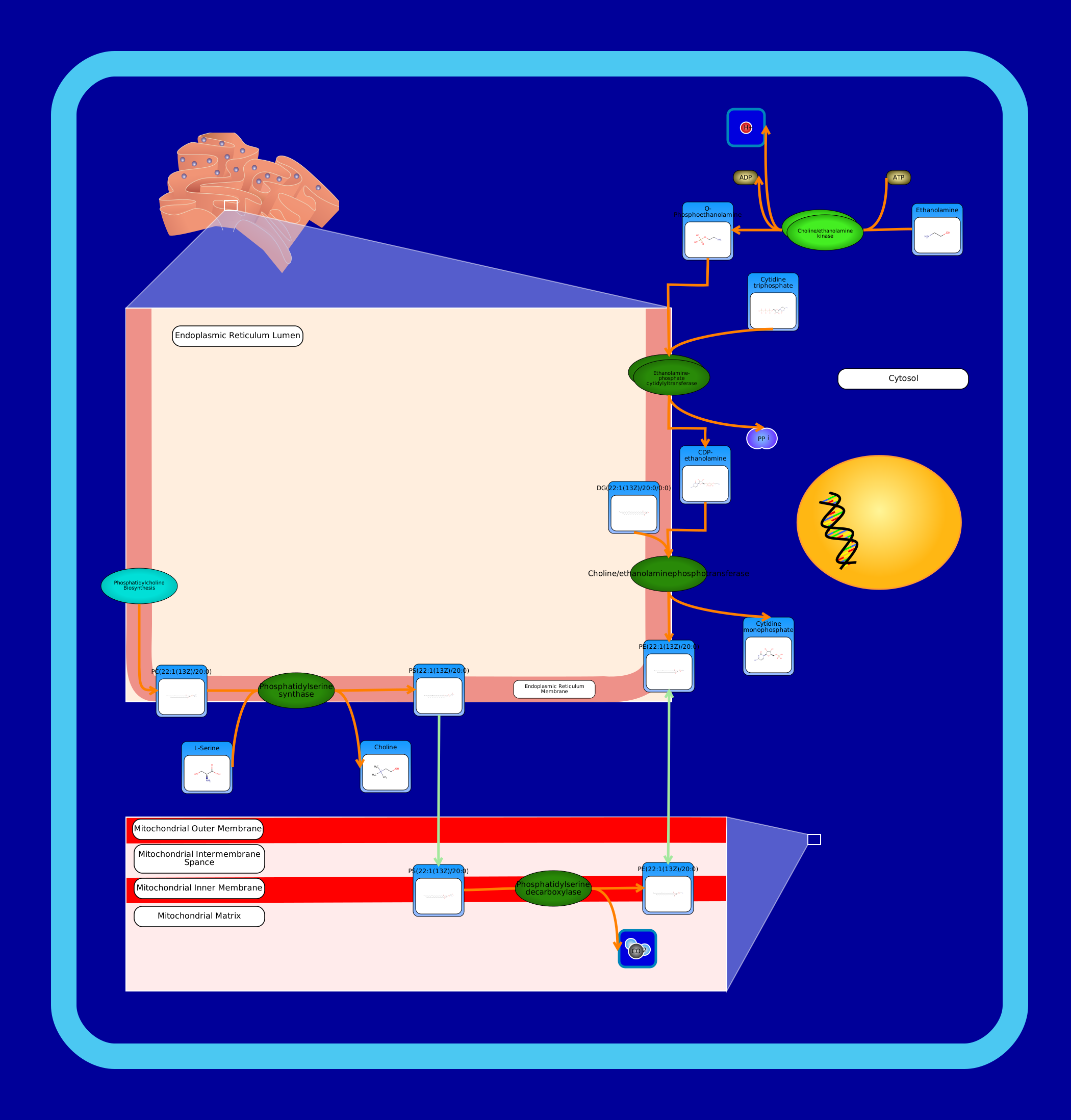 View Pathway
|
Phosphatidylethanolamine Biosynthesis PE(22:1(13Z)/20:0)Mus musculus
Phosphatidylethanolamines (PE) are a class of phospholipids that incorporate a phosphoric acid headgroup into a diacylglycerol backbone. They are the second most abundant phospholipid in eukaryotic cell membranes, and contrary to phosphatidylcholine, it is concentrated with phosphatidylserine in the cell membrane's inner leaflet. In Homo sapiens, there exist two phosphatidylethanolamine biosynthesis pathways. In the visualization, all enzymes that are dark green in colour are membrane-localized. The first pathway synthesizes phosphatidylethanolamine from ethanolamine via the Kennedy pathway. First, the cytosol-localized enzyme choline/ethanolamine kinase catalyzes the conversion of choline into phosphocholine. Second, choline-phosphate cytidylyltransferase, localized to the endoplasmic reticulum membrane, catalyzes the conversion of phosphocholine to CDP-choline. Last, choline/ethanolaminephosphotransferase catalyzes phosphatidylcholine biosynthesis from CDP-choline. It requires either magnesium or manganese ions as cofactors. Phosphatidylethanolamine is also synthesized from phosphatidylserine at the mitochondrial inner membrane by phosphatidylserine decarboxylase. Phosphatidylserine, itself, is synthesized using a base-exchange reaction with phosphatidylcholine. This reaction is catalyzed by phosphatidylserine synthase which is located in the endoplasmic reticulum membrane.
|
Metabolite
Metabolic
|
|
|
SMP0131600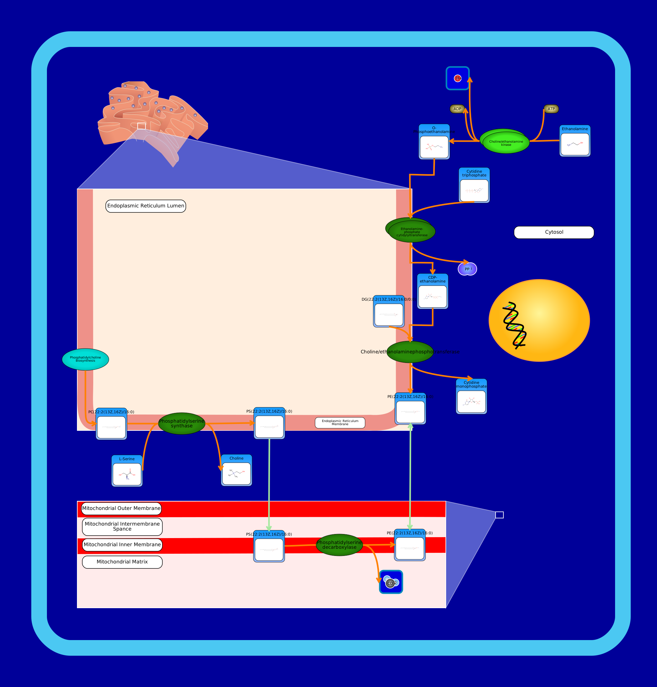 View Pathway
|
Phosphatidylethanolamine Biosynthesis PE(22:2(13Z,16Z)/16:0)Mus musculus
Phosphatidylethanolamines (PE) are a class of phospholipids that incorporate a phosphoric acid headgroup into a diacylglycerol backbone. They are the second most abundant phospholipid in eukaryotic cell membranes, and contrary to phosphatidylcholine, it is concentrated with phosphatidylserine in the cell membrane's inner leaflet. In Homo sapiens, there exist two phosphatidylethanolamine biosynthesis pathways. In the visualization, all enzymes that are dark green in colour are membrane-localized. The first pathway synthesizes phosphatidylethanolamine from ethanolamine via the Kennedy pathway. First, the cytosol-localized enzyme choline/ethanolamine kinase catalyzes the conversion of choline into phosphocholine. Second, choline-phosphate cytidylyltransferase, localized to the endoplasmic reticulum membrane, catalyzes the conversion of phosphocholine to CDP-choline. Last, choline/ethanolaminephosphotransferase catalyzes phosphatidylcholine biosynthesis from CDP-choline. It requires either magnesium or manganese ions as cofactors. Phosphatidylethanolamine is also synthesized from phosphatidylserine at the mitochondrial inner membrane by phosphatidylserine decarboxylase. Phosphatidylserine, itself, is synthesized using a base-exchange reaction with phosphatidylcholine. This reaction is catalyzed by phosphatidylserine synthase which is located in the endoplasmic reticulum membrane.
|
Metabolite
Metabolic
|
|
|
SMP0131581 View Pathway
|
Phosphatidylethanolamine Biosynthesis PE(22:1(13Z)/18:0)Mus musculus
Phosphatidylethanolamines (PE) are a class of phospholipids that incorporate a phosphoric acid headgroup into a diacylglycerol backbone. They are the second most abundant phospholipid in eukaryotic cell membranes, and contrary to phosphatidylcholine, it is concentrated with phosphatidylserine in the cell membrane's inner leaflet. In Homo sapiens, there exist two phosphatidylethanolamine biosynthesis pathways. In the visualization, all enzymes that are dark green in colour are membrane-localized. The first pathway synthesizes phosphatidylethanolamine from ethanolamine via the Kennedy pathway. First, the cytosol-localized enzyme choline/ethanolamine kinase catalyzes the conversion of choline into phosphocholine. Second, choline-phosphate cytidylyltransferase, localized to the endoplasmic reticulum membrane, catalyzes the conversion of phosphocholine to CDP-choline. Last, choline/ethanolaminephosphotransferase catalyzes phosphatidylcholine biosynthesis from CDP-choline. It requires either magnesium or manganese ions as cofactors. Phosphatidylethanolamine is also synthesized from phosphatidylserine at the mitochondrial inner membrane by phosphatidylserine decarboxylase. Phosphatidylserine, itself, is synthesized using a base-exchange reaction with phosphatidylcholine. This reaction is catalyzed by phosphatidylserine synthase which is located in the endoplasmic reticulum membrane.
|
Metabolite
Metabolic
|
|
|
SMP0131778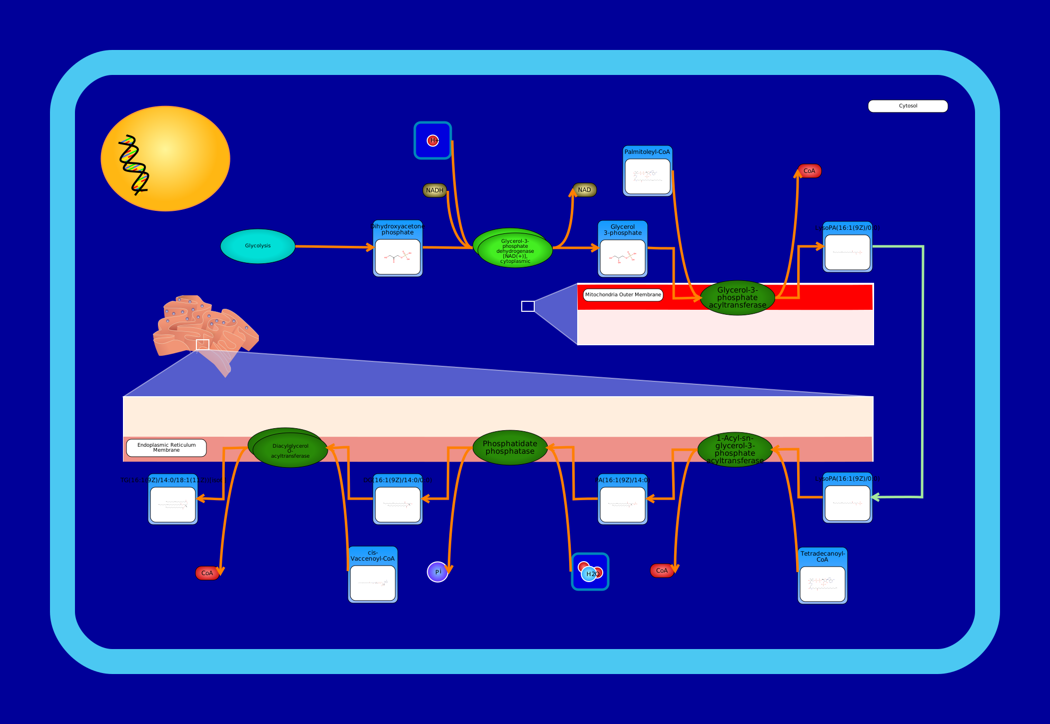 View Pathway
|
De Novo Triacylglycerol Biosynthesis TG(16:1(9Z)/14:0/18:1(11Z))Mus musculus
A triglyceride (TG, triacylglycerol, TAG, or triacylglyceride) is an ester derived from glycerol and three fatty acids. Triglycerides are the main constituents of body fat in humans and other animals, as well as vegetable fat. They are also present in the blood to enable the bidirectional transference of adipose fat and blood glucose from the liver, and are a major component of human skin oils. (Wikipedia) De novo biosynthesis of triglycerides is also known as the phosphatidic acid pathway, and it is mainly associated with the liver and adipose tissue. All membrane-localized enzymes are coloured dark green in the image. First, dihydroxyacetone phosphate (or glycerone phosphate) from glycolysis is used by the cytosolic enzyme glycerol-3-phosphate dehydrogenase [NAD(+)] to synthesize sn-glycerol 3-phosphate. Second, the mitochondrial outer membrane enzyme glycerol-3-phosphate acyltransferase esterifies an acyl-group to the sn-1 position of sn-glycerol 3-phosphate to form 1-acyl-sn-glycerol 3-phosphate (lysophosphatidic acid or LPA). The next three steps are localized to the endoplasmic reticulum membrane. The enzyme 1-acyl-sn-glycerol-3-phosphate acyltransferase converts LPA into phosphatidic acid (1,2-diacyl-sn-glycerol 3-phosphate) by esterifying an acyl-group to the sn-2 position of the glycerol backbone. Next, magnesium-dependent phosphatidate phosphatase catalyzes the conversion of phosphatidic acid into diacylglycerol. Last, the enzyme diacylglycerol O-acyltransferase synthesizes triacylglycerol from diacylglycerol and a fatty acyl-CoA.
|
Metabolite
Metabolic
|
||
SMP0131797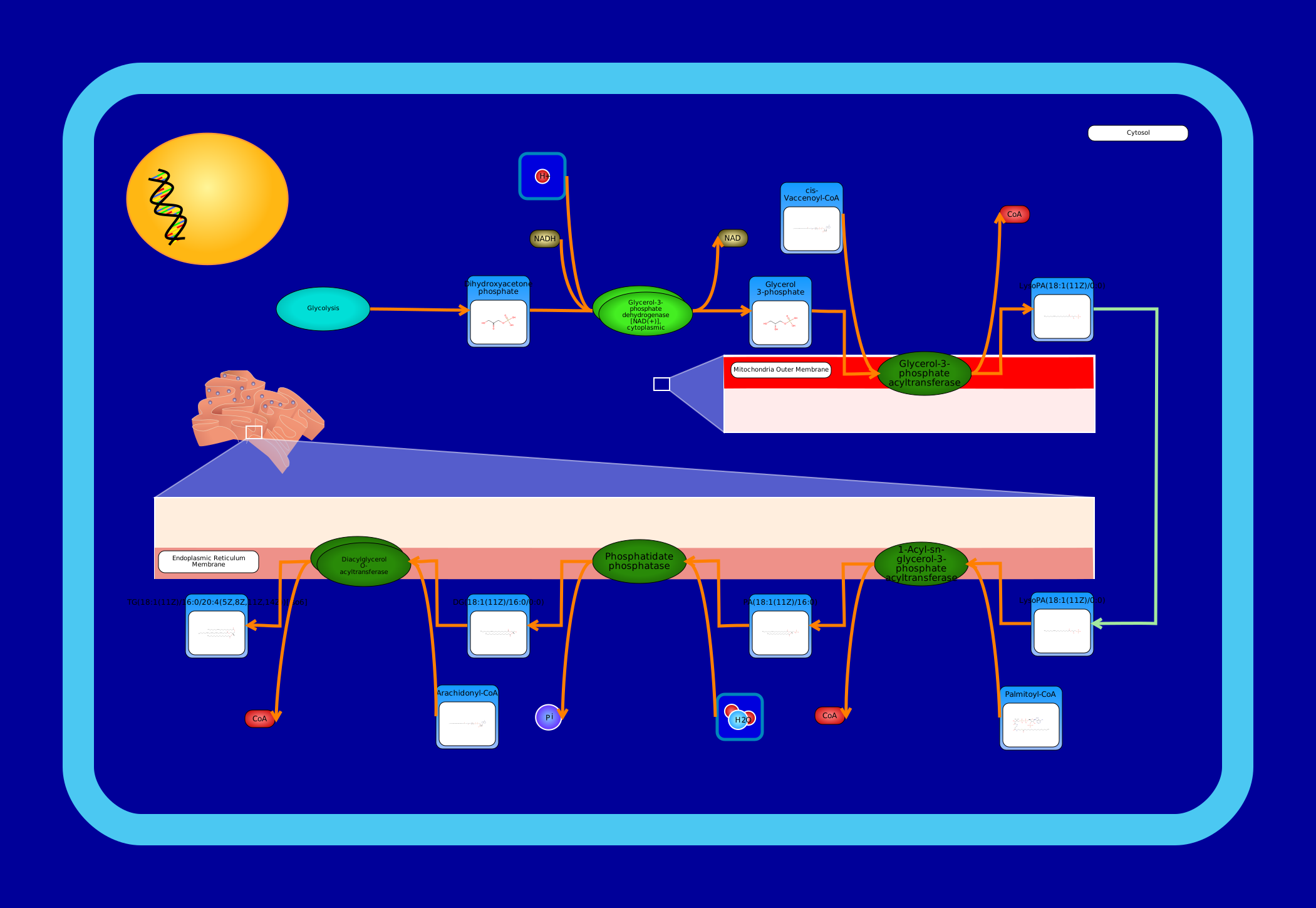 View Pathway
|
De Novo Triacylglycerol Biosynthesis TG(18:1(11Z)/16:0/20:4(5Z,8Z,11Z,14Z))Mus musculus
A triglyceride (TG, triacylglycerol, TAG, or triacylglyceride) is an ester derived from glycerol and three fatty acids. Triglycerides are the main constituents of body fat in humans and other animals, as well as vegetable fat. They are also present in the blood to enable the bidirectional transference of adipose fat and blood glucose from the liver, and are a major component of human skin oils. (Wikipedia) De novo biosynthesis of triglycerides is also known as the phosphatidic acid pathway, and it is mainly associated with the liver and adipose tissue. All membrane-localized enzymes are coloured dark green in the image. First, dihydroxyacetone phosphate (or glycerone phosphate) from glycolysis is used by the cytosolic enzyme glycerol-3-phosphate dehydrogenase [NAD(+)] to synthesize sn-glycerol 3-phosphate. Second, the mitochondrial outer membrane enzyme glycerol-3-phosphate acyltransferase esterifies an acyl-group to the sn-1 position of sn-glycerol 3-phosphate to form 1-acyl-sn-glycerol 3-phosphate (lysophosphatidic acid or LPA). The next three steps are localized to the endoplasmic reticulum membrane. The enzyme 1-acyl-sn-glycerol-3-phosphate acyltransferase converts LPA into phosphatidic acid (1,2-diacyl-sn-glycerol 3-phosphate) by esterifying an acyl-group to the sn-2 position of the glycerol backbone. Next, magnesium-dependent phosphatidate phosphatase catalyzes the conversion of phosphatidic acid into diacylglycerol. Last, the enzyme diacylglycerol O-acyltransferase synthesizes triacylglycerol from diacylglycerol and a fatty acyl-CoA.
|
Metabolite
Metabolic
|
||
SMP0131773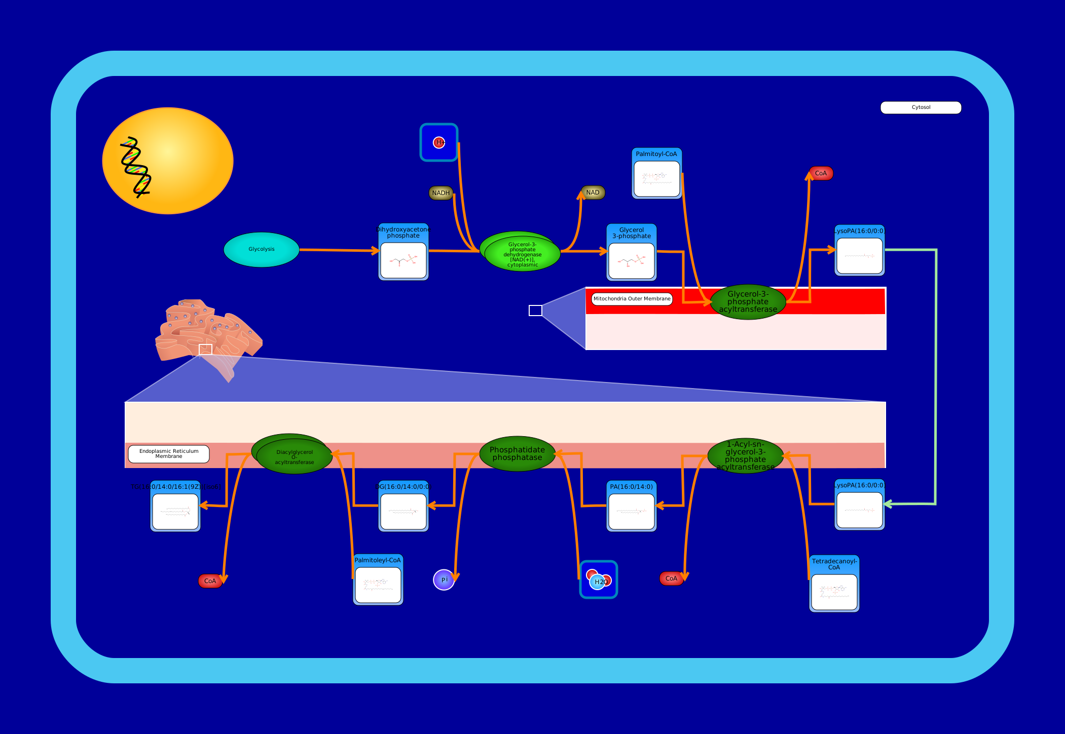 View Pathway
|
De Novo Triacylglycerol Biosynthesis TG(16:0/14:0/16:1(9Z))Mus musculus
A triglyceride (TG, triacylglycerol, TAG, or triacylglyceride) is an ester derived from glycerol and three fatty acids. Triglycerides are the main constituents of body fat in humans and other animals, as well as vegetable fat. They are also present in the blood to enable the bidirectional transference of adipose fat and blood glucose from the liver, and are a major component of human skin oils. (Wikipedia) De novo biosynthesis of triglycerides is also known as the phosphatidic acid pathway, and it is mainly associated with the liver and adipose tissue. All membrane-localized enzymes are coloured dark green in the image. First, dihydroxyacetone phosphate (or glycerone phosphate) from glycolysis is used by the cytosolic enzyme glycerol-3-phosphate dehydrogenase [NAD(+)] to synthesize sn-glycerol 3-phosphate. Second, the mitochondrial outer membrane enzyme glycerol-3-phosphate acyltransferase esterifies an acyl-group to the sn-1 position of sn-glycerol 3-phosphate to form 1-acyl-sn-glycerol 3-phosphate (lysophosphatidic acid or LPA). The next three steps are localized to the endoplasmic reticulum membrane. The enzyme 1-acyl-sn-glycerol-3-phosphate acyltransferase converts LPA into phosphatidic acid (1,2-diacyl-sn-glycerol 3-phosphate) by esterifying an acyl-group to the sn-2 position of the glycerol backbone. Next, magnesium-dependent phosphatidate phosphatase catalyzes the conversion of phosphatidic acid into diacylglycerol. Last, the enzyme diacylglycerol O-acyltransferase synthesizes triacylglycerol from diacylglycerol and a fatty acyl-CoA.
|
Metabolite
Metabolic
|
||
SMP0131792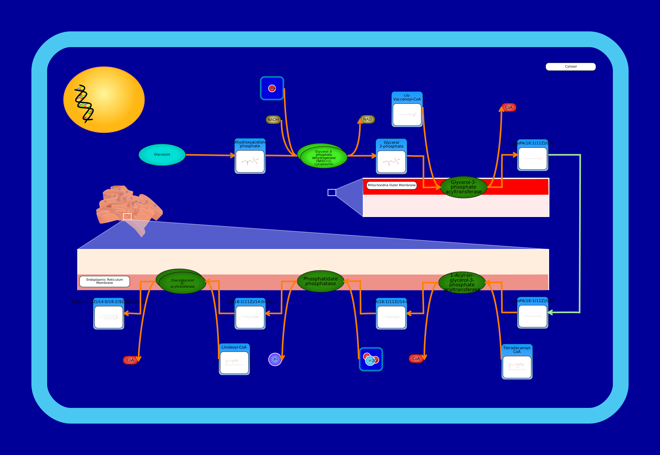 View Pathway
|
De Novo Triacylglycerol Biosynthesis TG(18:1(11Z)/14:0/18:2(9Z,12Z))Mus musculus
A triglyceride (TG, triacylglycerol, TAG, or triacylglyceride) is an ester derived from glycerol and three fatty acids. Triglycerides are the main constituents of body fat in humans and other animals, as well as vegetable fat. They are also present in the blood to enable the bidirectional transference of adipose fat and blood glucose from the liver, and are a major component of human skin oils. (Wikipedia) De novo biosynthesis of triglycerides is also known as the phosphatidic acid pathway, and it is mainly associated with the liver and adipose tissue. All membrane-localized enzymes are coloured dark green in the image. First, dihydroxyacetone phosphate (or glycerone phosphate) from glycolysis is used by the cytosolic enzyme glycerol-3-phosphate dehydrogenase [NAD(+)] to synthesize sn-glycerol 3-phosphate. Second, the mitochondrial outer membrane enzyme glycerol-3-phosphate acyltransferase esterifies an acyl-group to the sn-1 position of sn-glycerol 3-phosphate to form 1-acyl-sn-glycerol 3-phosphate (lysophosphatidic acid or LPA). The next three steps are localized to the endoplasmic reticulum membrane. The enzyme 1-acyl-sn-glycerol-3-phosphate acyltransferase converts LPA into phosphatidic acid (1,2-diacyl-sn-glycerol 3-phosphate) by esterifying an acyl-group to the sn-2 position of the glycerol backbone. Next, magnesium-dependent phosphatidate phosphatase catalyzes the conversion of phosphatidic acid into diacylglycerol. Last, the enzyme diacylglycerol O-acyltransferase synthesizes triacylglycerol from diacylglycerol and a fatty acyl-CoA.
|
Metabolite
Metabolic
|
Showing 541201 -
541210 of 541248 pathways