
Browsing Pathways
Showing 541211 -
541220 of 605359 pathways
| PathBank ID | Pathway Name and Description | Pathway Class | Chemical Compounds | Proteins |
|---|---|---|---|---|
SMP0131790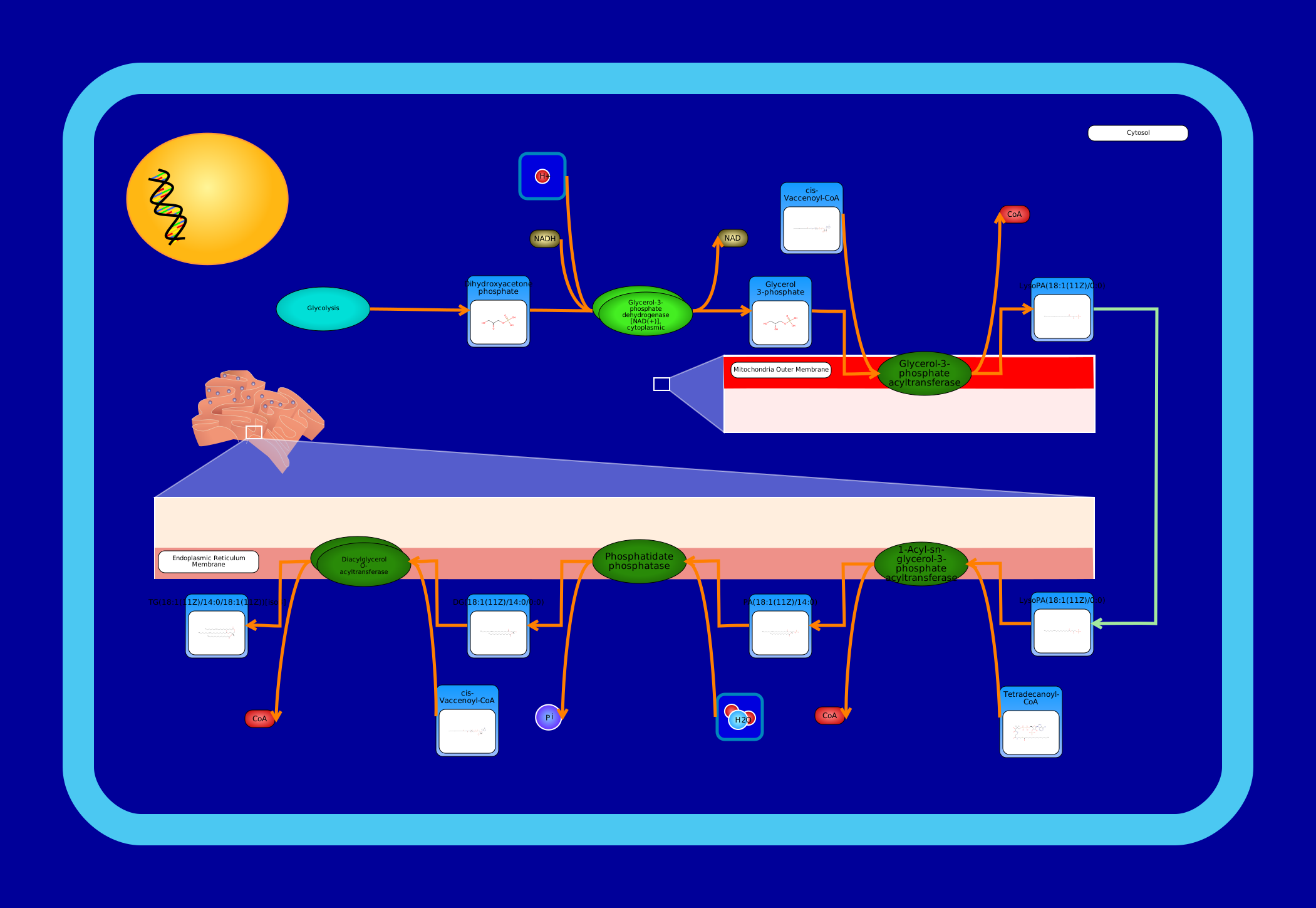 View Pathway
|
De Novo Triacylglycerol Biosynthesis TG(18:1(11Z)/14:0/18:1(11Z))Mus musculus
A triglyceride (TG, triacylglycerol, TAG, or triacylglyceride) is an ester derived from glycerol and three fatty acids. Triglycerides are the main constituents of body fat in humans and other animals, as well as vegetable fat. They are also present in the blood to enable the bidirectional transference of adipose fat and blood glucose from the liver, and are a major component of human skin oils. (Wikipedia) De novo biosynthesis of triglycerides is also known as the phosphatidic acid pathway, and it is mainly associated with the liver and adipose tissue. All membrane-localized enzymes are coloured dark green in the image. First, dihydroxyacetone phosphate (or glycerone phosphate) from glycolysis is used by the cytosolic enzyme glycerol-3-phosphate dehydrogenase [NAD(+)] to synthesize sn-glycerol 3-phosphate. Second, the mitochondrial outer membrane enzyme glycerol-3-phosphate acyltransferase esterifies an acyl-group to the sn-1 position of sn-glycerol 3-phosphate to form 1-acyl-sn-glycerol 3-phosphate (lysophosphatidic acid or LPA). The next three steps are localized to the endoplasmic reticulum membrane. The enzyme 1-acyl-sn-glycerol-3-phosphate acyltransferase converts LPA into phosphatidic acid (1,2-diacyl-sn-glycerol 3-phosphate) by esterifying an acyl-group to the sn-2 position of the glycerol backbone. Next, magnesium-dependent phosphatidate phosphatase catalyzes the conversion of phosphatidic acid into diacylglycerol. Last, the enzyme diacylglycerol O-acyltransferase synthesizes triacylglycerol from diacylglycerol and a fatty acyl-CoA.
|
Metabolite
Metabolic
|
||
SMP0131759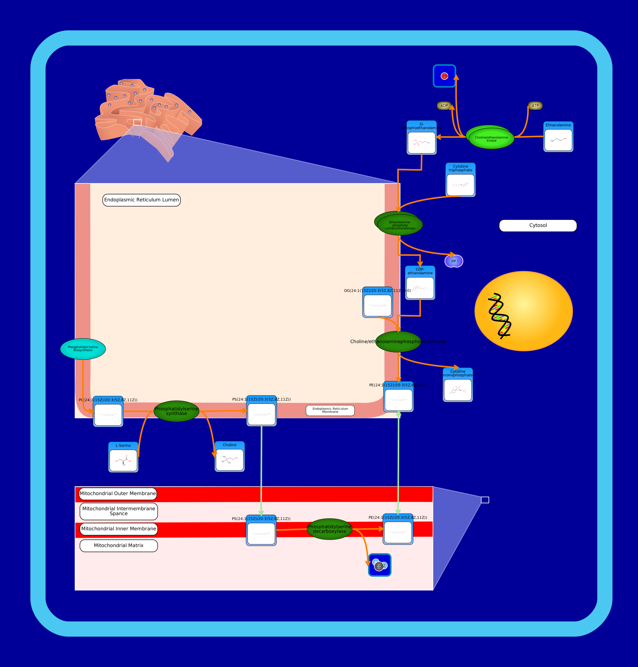 View Pathway
|
Phosphatidylethanolamine Biosynthesis PE(24:1(15Z)/20:3(5Z,8Z,11Z))Mus musculus
Phosphatidylethanolamines (PE) are a class of phospholipids that incorporate a phosphoric acid headgroup into a diacylglycerol backbone. They are the second most abundant phospholipid in eukaryotic cell membranes, and contrary to phosphatidylcholine, it is concentrated with phosphatidylserine in the cell membrane's inner leaflet. In Homo sapiens, there exist two phosphatidylethanolamine biosynthesis pathways. In the visualization, all enzymes that are dark green in colour are membrane-localized. The first pathway synthesizes phosphatidylethanolamine from ethanolamine via the Kennedy pathway. First, the cytosol-localized enzyme choline/ethanolamine kinase catalyzes the conversion of choline into phosphocholine. Second, choline-phosphate cytidylyltransferase, localized to the endoplasmic reticulum membrane, catalyzes the conversion of phosphocholine to CDP-choline. Last, choline/ethanolaminephosphotransferase catalyzes phosphatidylcholine biosynthesis from CDP-choline. It requires either magnesium or manganese ions as cofactors. Phosphatidylethanolamine is also synthesized from phosphatidylserine at the mitochondrial inner membrane by phosphatidylserine decarboxylase. Phosphatidylserine, itself, is synthesized using a base-exchange reaction with phosphatidylcholine. This reaction is catalyzed by phosphatidylserine synthase which is located in the endoplasmic reticulum membrane.
|
Metabolite
Metabolic
|
|
|
SMP0131766 View Pathway
|
Phosphatidylethanolamine Biosynthesis PE(24:1(15Z)/22:2(13Z,16Z))Mus musculus
Phosphatidylethanolamines (PE) are a class of phospholipids that incorporate a phosphoric acid headgroup into a diacylglycerol backbone. They are the second most abundant phospholipid in eukaryotic cell membranes, and contrary to phosphatidylcholine, it is concentrated with phosphatidylserine in the cell membrane's inner leaflet. In Homo sapiens, there exist two phosphatidylethanolamine biosynthesis pathways. In the visualization, all enzymes that are dark green in colour are membrane-localized. The first pathway synthesizes phosphatidylethanolamine from ethanolamine via the Kennedy pathway. First, the cytosol-localized enzyme choline/ethanolamine kinase catalyzes the conversion of choline into phosphocholine. Second, choline-phosphate cytidylyltransferase, localized to the endoplasmic reticulum membrane, catalyzes the conversion of phosphocholine to CDP-choline. Last, choline/ethanolaminephosphotransferase catalyzes phosphatidylcholine biosynthesis from CDP-choline. It requires either magnesium or manganese ions as cofactors. Phosphatidylethanolamine is also synthesized from phosphatidylserine at the mitochondrial inner membrane by phosphatidylserine decarboxylase. Phosphatidylserine, itself, is synthesized using a base-exchange reaction with phosphatidylcholine. This reaction is catalyzed by phosphatidylserine synthase which is located in the endoplasmic reticulum membrane.
|
Metabolite
Metabolic
|
|
|
SMP0131785 View Pathway
|
De Novo Triacylglycerol Biosynthesis TG(18:0/14:0/18:1(11Z))Mus musculus
A triglyceride (TG, triacylglycerol, TAG, or triacylglyceride) is an ester derived from glycerol and three fatty acids. Triglycerides are the main constituents of body fat in humans and other animals, as well as vegetable fat. They are also present in the blood to enable the bidirectional transference of adipose fat and blood glucose from the liver, and are a major component of human skin oils. (Wikipedia) De novo biosynthesis of triglycerides is also known as the phosphatidic acid pathway, and it is mainly associated with the liver and adipose tissue. All membrane-localized enzymes are coloured dark green in the image. First, dihydroxyacetone phosphate (or glycerone phosphate) from glycolysis is used by the cytosolic enzyme glycerol-3-phosphate dehydrogenase [NAD(+)] to synthesize sn-glycerol 3-phosphate. Second, the mitochondrial outer membrane enzyme glycerol-3-phosphate acyltransferase esterifies an acyl-group to the sn-1 position of sn-glycerol 3-phosphate to form 1-acyl-sn-glycerol 3-phosphate (lysophosphatidic acid or LPA). The next three steps are localized to the endoplasmic reticulum membrane. The enzyme 1-acyl-sn-glycerol-3-phosphate acyltransferase converts LPA into phosphatidic acid (1,2-diacyl-sn-glycerol 3-phosphate) by esterifying an acyl-group to the sn-2 position of the glycerol backbone. Next, magnesium-dependent phosphatidate phosphatase catalyzes the conversion of phosphatidic acid into diacylglycerol. Last, the enzyme diacylglycerol O-acyltransferase synthesizes triacylglycerol from diacylglycerol and a fatty acyl-CoA.
|
Metabolite
Metabolic
|
||
SMP0131771 View Pathway
|
Phosphatidylethanolamine Biosynthesis PE(24:1(15Z)/24:0)Mus musculus
Phosphatidylethanolamines (PE) are a class of phospholipids that incorporate a phosphoric acid headgroup into a diacylglycerol backbone. They are the second most abundant phospholipid in eukaryotic cell membranes, and contrary to phosphatidylcholine, it is concentrated with phosphatidylserine in the cell membrane's inner leaflet. In Homo sapiens, there exist two phosphatidylethanolamine biosynthesis pathways. In the visualization, all enzymes that are dark green in colour are membrane-localized. The first pathway synthesizes phosphatidylethanolamine from ethanolamine via the Kennedy pathway. First, the cytosol-localized enzyme choline/ethanolamine kinase catalyzes the conversion of choline into phosphocholine. Second, choline-phosphate cytidylyltransferase, localized to the endoplasmic reticulum membrane, catalyzes the conversion of phosphocholine to CDP-choline. Last, choline/ethanolaminephosphotransferase catalyzes phosphatidylcholine biosynthesis from CDP-choline. It requires either magnesium or manganese ions as cofactors. Phosphatidylethanolamine is also synthesized from phosphatidylserine at the mitochondrial inner membrane by phosphatidylserine decarboxylase. Phosphatidylserine, itself, is synthesized using a base-exchange reaction with phosphatidylcholine. This reaction is catalyzed by phosphatidylserine synthase which is located in the endoplasmic reticulum membrane.
|
Metabolite
Metabolic
|
|
|
SMP0121111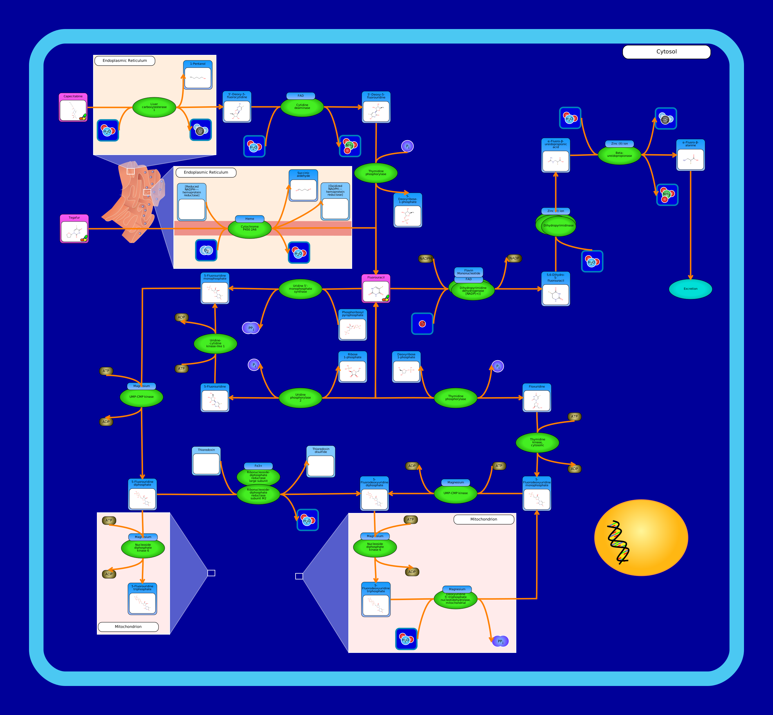 View Pathway
|
Fluorouracil MetabolismHomo sapiens
Fluorouracil, sold as Adrucil, Carac, Efudex, Efudix and others, is a medication used to treat various forms of cancer. It consists of a fluorine atom on the 5th carbon of a uracil molecule, and is treated similarly to the uracil during metabolism by the body, but the fluorouracil tends to be absorbed more readily by tumor cells than healthy cells, allowing it to target cancer cells.
Capecitabine is one of the prodrugs that can be metabolized into fluorouracil. First, it is converted to 5'-deoxy-5-fluorocytidine by liver carboxylesterase 1 in the endoplasmic reticulum, and then by cytidine deaminase into 5'-deoxy-5-fluorouridine. Finally, it is converted into fluorouracil by thymidine, which removes the 5'-deoxyribose-1-phosphate from it.
Tegafur is another prodrug that may be converted to fluorouracil, this time by cytochrome P450 2A6 in the endoplasmic reticulum membrane.
From this point, fluorouracil can be converted to 5,6-dihydro-5-fluorouracil by dihydropyrimidine dehydrogenase, which adds a hydrogen ion to it. The 5,6-dihydro-5-fluorouracil can then have a water molecule added by dihydropyrimidinase, forming alpha-fluoro-beta-ureidopropionic acid. Finally, this is converted to alpha-fluoro-beta-alanine by beta-ureidopropionase, which is an end product of the pathway, and is then excreted.
Fluorouracil can also be converted to and from 5-fluorouridine by uridine phosphorylase 2, which is then converted to 5-fluorouridine monophosphate by urudine-cytidine kinase-like 1. 5-fluorouridine monophosphate is also formed from fluorouracil via catalysis by uridine 5'-monophosphate synthase. Regardless of the pathway through which it is created, 5-fluorouridine monophosphate is converted to 5-fluorouridine diphosphate by UMP-CMP kinase, which adds a phosphate group to it. Whithin the mitochondria, mucleoside diphosphate kinase 6 adds one final phosphate group to it, forming 5-fluorouridine triphosphate, another end product of the pathway.
If it does not enter the mitochondria, 5-fluorouridine diphosphate can instead be converted to 5-fluorodeoxyuridine diphosphate by the ribonucleoside-diphosphate reductase complex.
Finally, fluorouracil may be converted to and from floxuridine by thymidine phosphorylase, which then is converted to 5-fluorodeoxyuridine monophosphate by cytosolic thymidine kinase. This molecule also forms 5-fluorodeoxyuridine diphosphate via UMP-CMP kinase, bringing these two branches of the pathway together. Regardless of its origin, 5-fluorodeoxyuridine diphosphate can be converted to 5-fluorodeoxyuridine triphosphate by nucleoside diphosphate kinase 6 in the mitochondria. It may stop there, or be converted back to 5-fluorodeoxyuridine monophosphate by deoxyuridine 5'-triphosphate nucleotidohydrolase, also in the mitochondria.
|
Metabolite
Metabolic
|
|
|
SMP0121104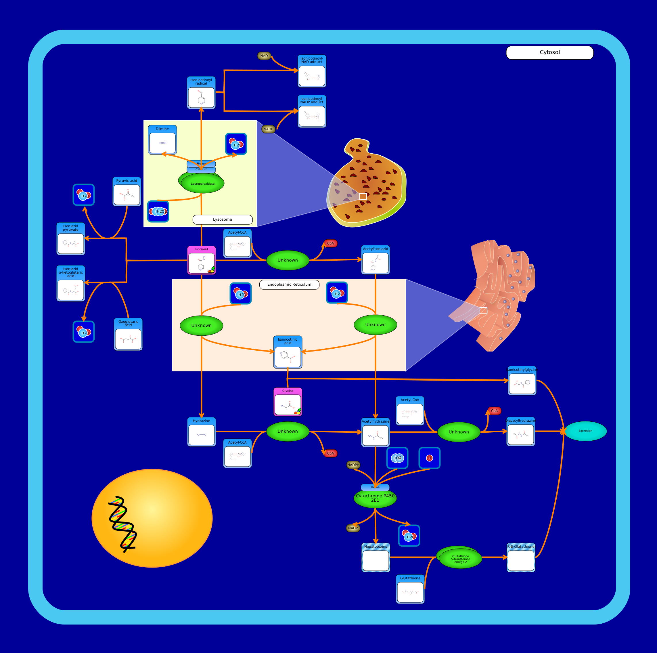 View Pathway
|
Isoniazid MetabolismBos taurus
Isoniazid is an antibiotic drug used to treat tunerculosis, as well as other types of mycobacteria. Through a currently unknown reaction that may be spontaneous or enzymatic, pyruvic acid or oxoglutaric acid can undergo a dehydration reaction with isoniazid, forming isoniazid pyruvate or isoniazid alpha-ketoglutaric acid. Isoniazid may also react with hydrogen peroxide in the lysosome, forming an isonicotinoyl radical catalyzed by myeloperoxidase. The isonicotinoyl radical can then have either NAD or NADP added in a non-enzymatic reaction, forming isonicotinoyl-NAD and NADP adducts. Isoniazid can have an acetyl group added to it by arylamine N-acetyltransferase 2, fvorming acetylisoniazid. This can then enter the endoplasmic reticulum and, with the addition of a water molecule, can form isonicotinic acid and acetylhydrazine. Isoniazid can also be converted to hydrazine and isonicotinic acid via the same reaction, and the hydrazine can have an acetyl group added to it by arylamine N-acetyltransferase 2 in order to form acetylhydrazine. Acetylhydrazine can have another acetyl group added to it by arylamine N-acetyltransferase 2 to form diacetylhydrazine which is then excreted. It can alternatively be processed by cytochrome P450 2E1 into hepatotoxins, which are then joined to glutatione by glutatione S-transferase omega-2 to form R-S-glutatione, which is then excreted. Finally, isonicotinic acid can react with a glycine in an unclear reaction, potentially requiring ATP and coenzyme A and forming an intermediate, producing isonicotinylglycine, which is also excreted.
|
Metabolite
Metabolic
|
|
|
SMP0121086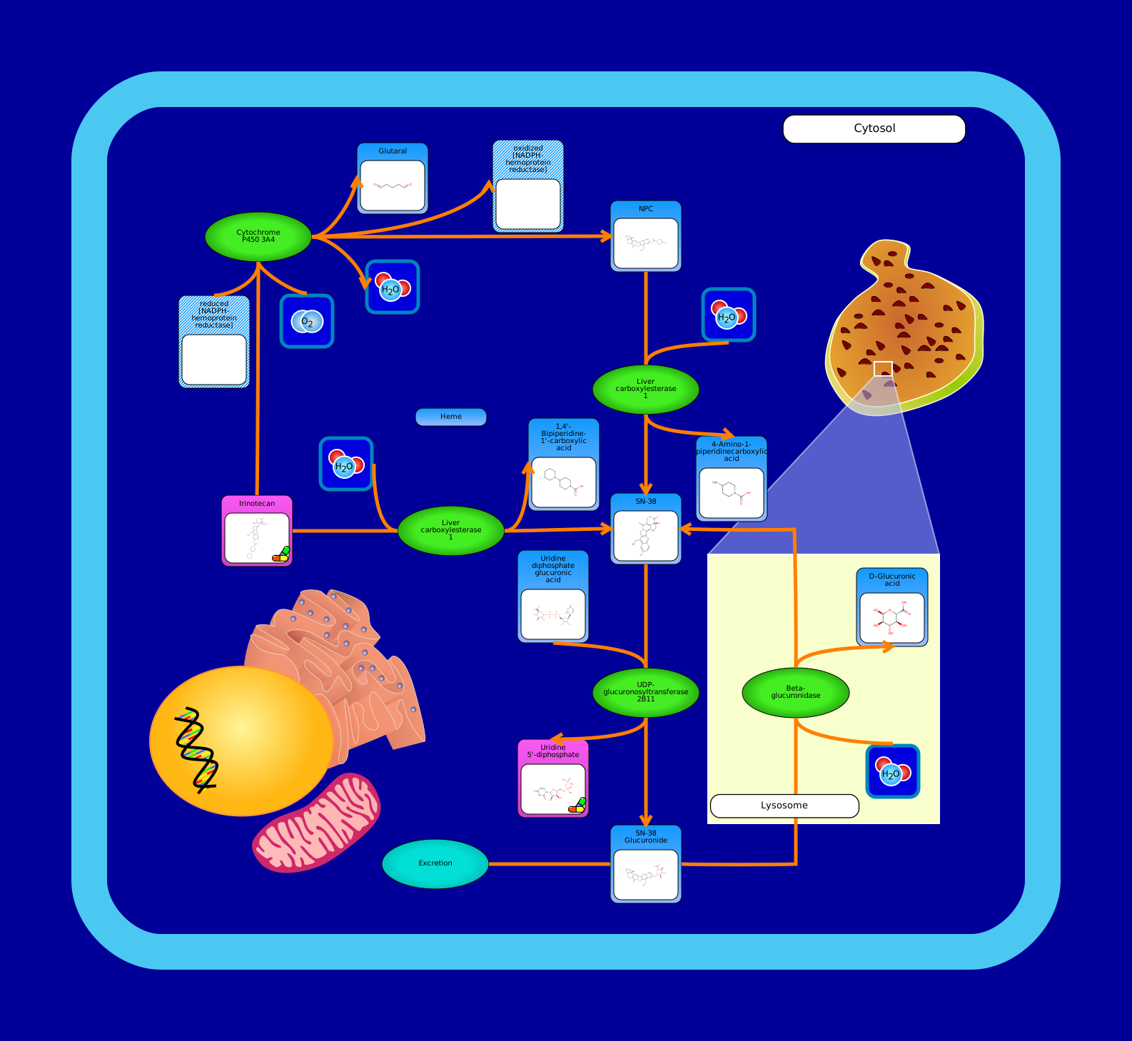 View Pathway
|
Irinotecan MetabolismHomo sapiens
Irinotecan, branded as Camptosar, Campto, Onivyde and others, is a cancer medication used to treat colon and small cell lung cancers, alone or with other drugs.
Irinotecan can be processed by the cytochrome P450 3A4 enzyme, producing both the side product glutaral, as well as a compound called NPC. the NPC can then be catalyzed by liver carboxylesterase 1 to form 7-ethyl-10-hydroxy-camptothecin, or SN-38. Alternatively, irinotecan can directly form SN-38 via catalysis by liver carboxylesterase 1.
After its formation, SN-38 is converted to SN-38 glucuronide by UDP-glucuronosyltransferase 2B11. This can then be converted back to SN-38 in the lysosome by beta-glucuronidase, or can be excreted as the end product of the pathway.
|
Metabolite
Metabolic
|
|
|
SMP0121097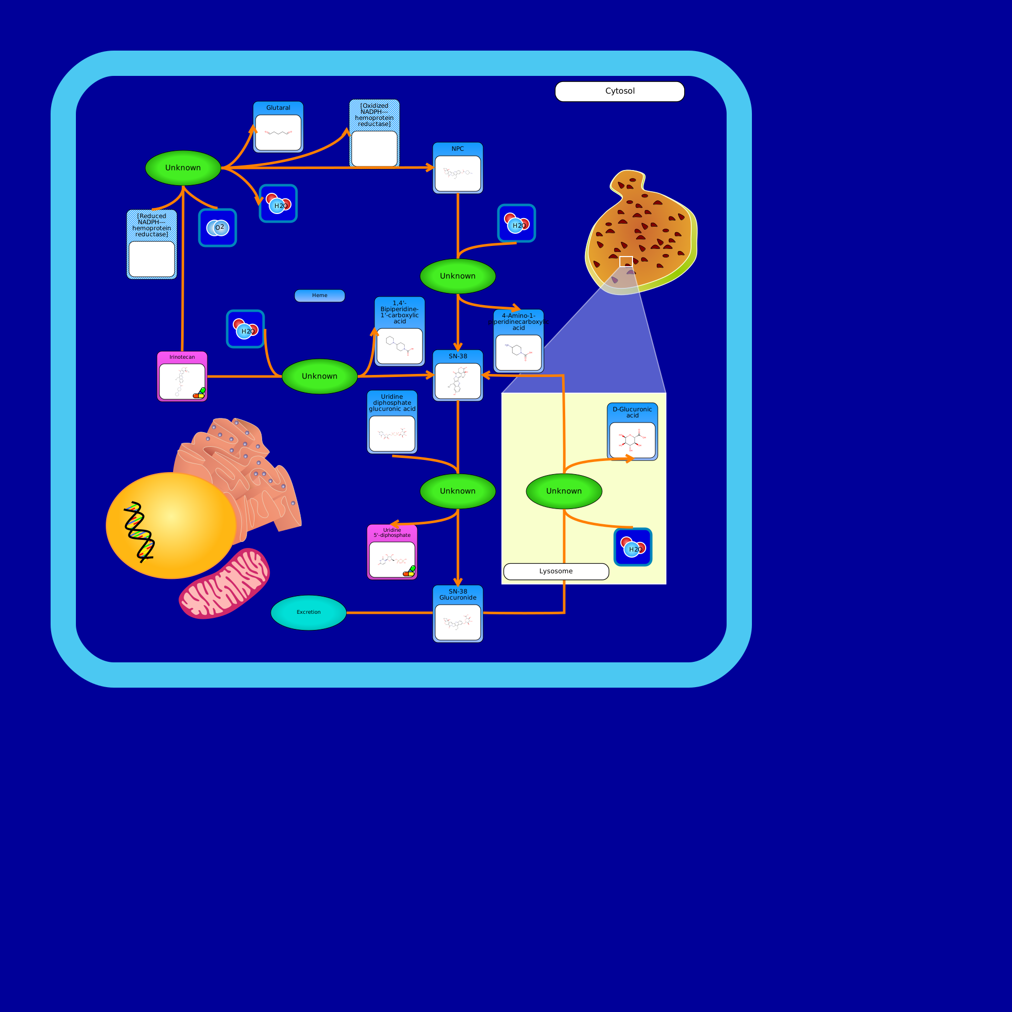 View Pathway
|
Irinotecan MetabolismDanio rerio
|
Metabolite
Metabolic
|
||
SMP0121109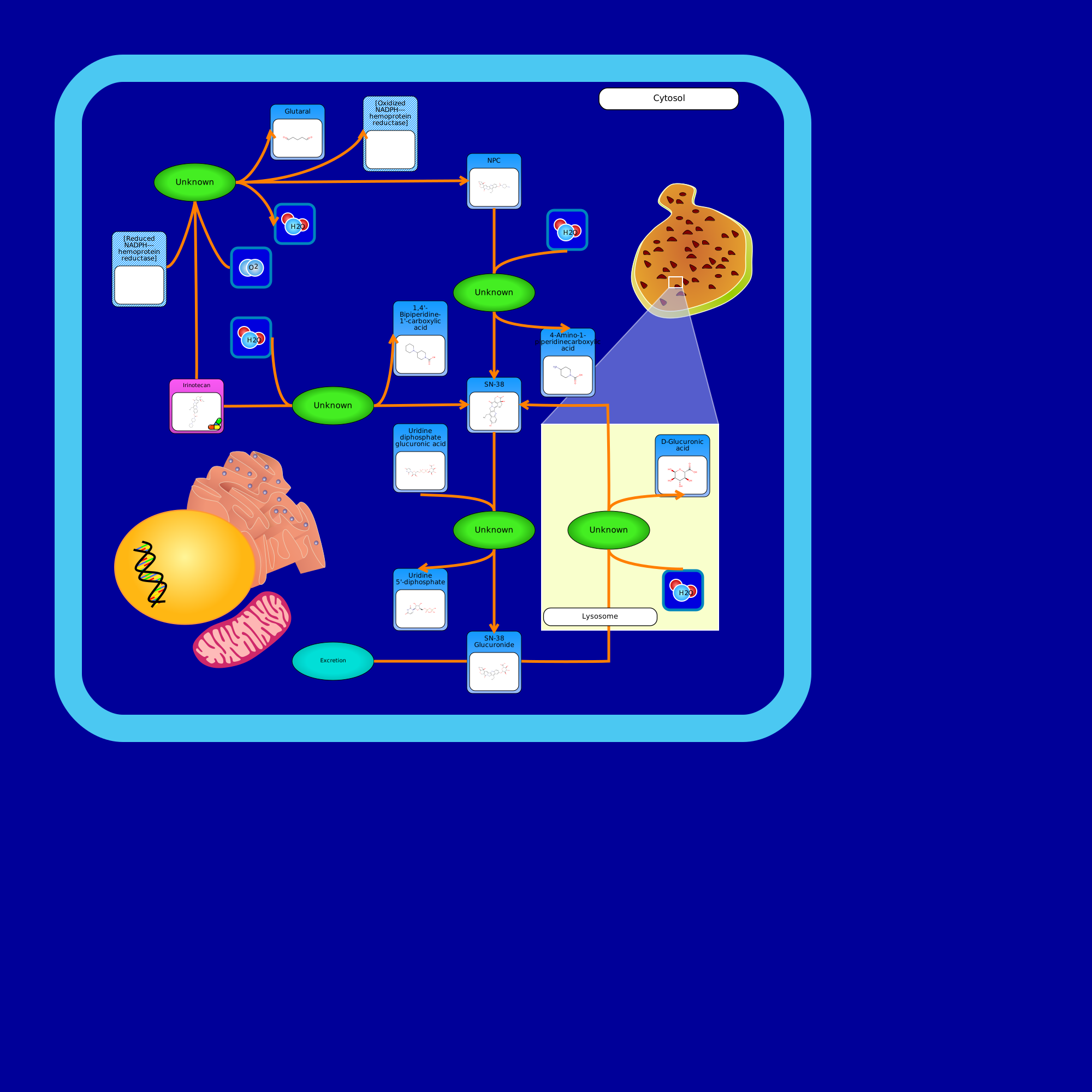 View Pathway
|
Irinotecan MetabolismCaenorhabditis elegans
|
Metabolite
Metabolic
|
Showing 541211 -
541220 of 541248 pathways