
Browsing Pathways
Showing 21 -
30 of 605359 pathways
| PathBank ID | Pathway Name and Description | Pathway Class | Chemical Compounds | Proteins |
|---|---|---|---|---|
SMP0121063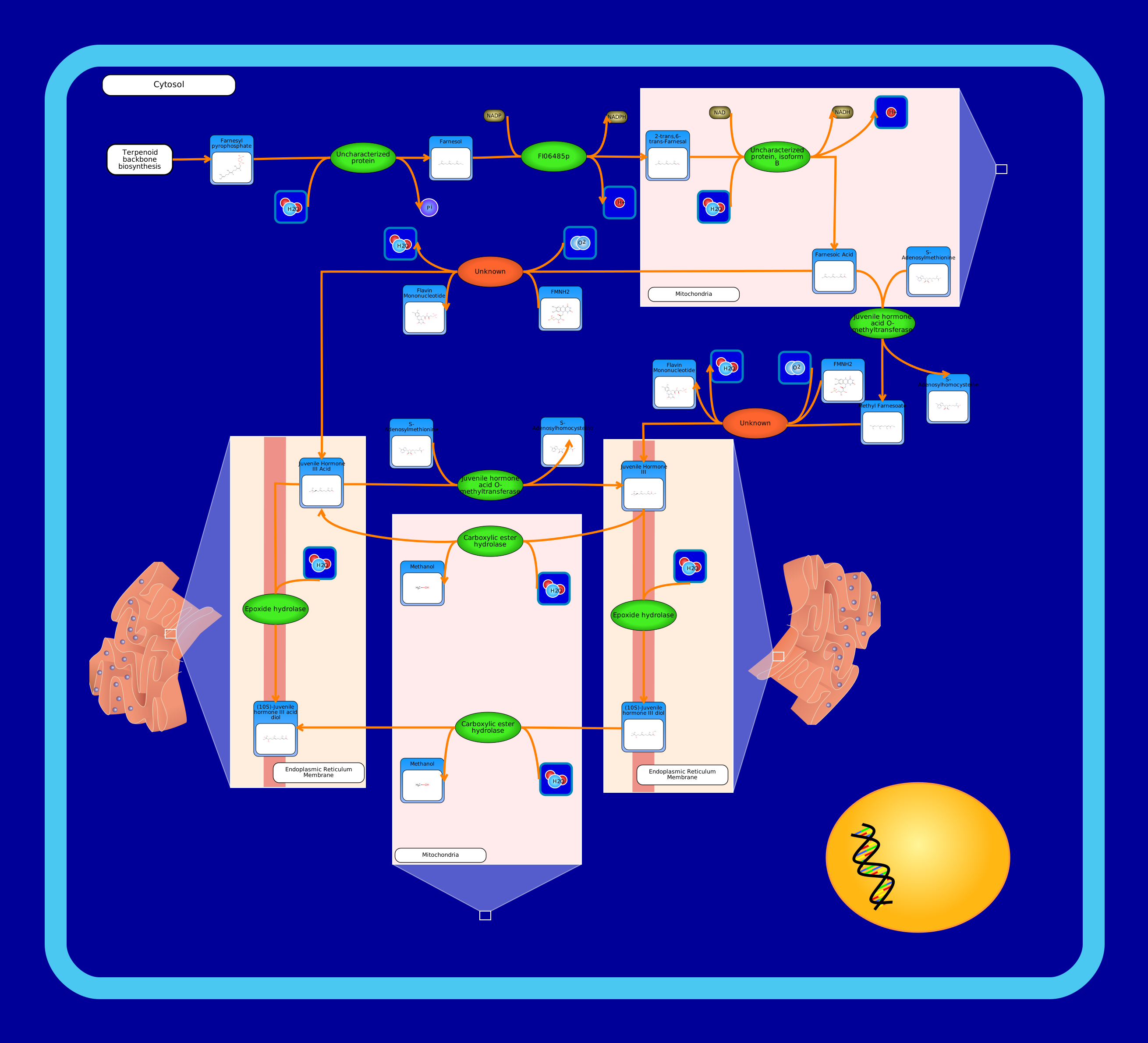 View Pathway
|
Juvenile Hormone SynthesisDrosophila melanogaster
Juvenile hormones in insects are important for their growth before their adulthood, preventing metamorphosis if they undergo one. In Drosophila, only juvenile hormone III has been identified, while others exist in butterflies and moths.
Synthesis of various forms of juvenile hormone III (JH III) start with farnesyl diphosphate interacting with an uncharacterized phosphatase protein, forming farnesol. Farnesol then interacts with NADP+ dependent farensol dehydrogenase, which removes a hydrogen ion from the hydroxyl group in order to form farnesal. Farnesal then enters the mitochondria and interacts with another uncharacterized aldehyde dehydrogenase which allows it to form farnesoic acid. Farnesoic acid can then interact with an unknown protein, similar to farnesoate epoxidase in Bombyx mori, in order to form juvenile hormone III acid (JH III acid). JH III acid can then interact with epoxide hydrolase in the membrane of the endoplasmic reticulum, forming the final product of this pathway, juvenile hormone III acid diol (JH III acid diol). It can also interact with juvenile hormone acid O-methyltransferase in order to form JH III, which is used in another set of reactions in this pathway.
If farnesoic acid does not interact with the unknown protein, it may interact with juvenile hormone acid O-methyltransferase to form methyl farnesoate. Methyl farnesoate can then interact with a different unknown protein similar, to methyl farnesoate epoxidase in Diploptera punctata, in order to form JH III. In the mitochondria, JH III can interact with carboxylic ester hydrolase in order to form JH III acid, which then can form the final product, or form JH III again. Alternately, JH III can interact with epoxide hydrolase in the membrane of the endoplasmic reticulum, forming juvenile hormone III diol. This product then interacts with carboxylic ester hydrolase in the mitochondria, forming JH III acid diol, again, the end product of this pathway.
|
Metabolite
Metabolic
|
|
|
SMP0121068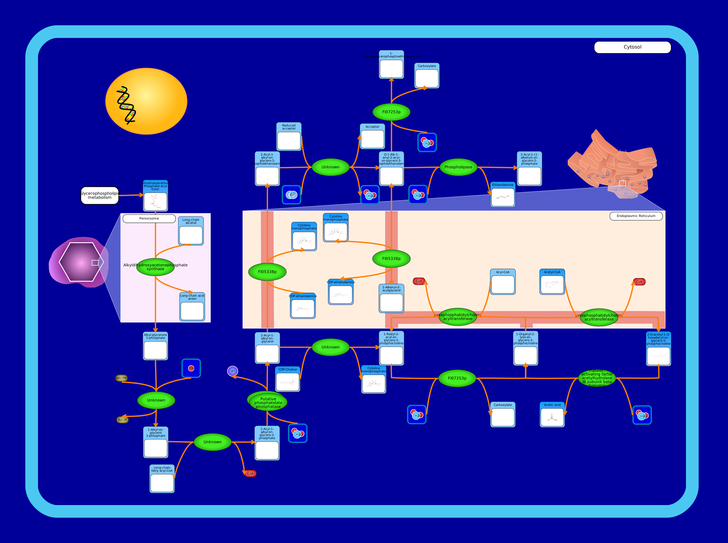 View Pathway
|
Ether Lipid MetabolismDrosophila melanogaster
Ether lipids are typically glycerophospholipids where the glycerol backbone has lipids attached by both an ether bond at the sn-1 position and an acyl group at the sn-2 position.
This pathway starts with dihydroxyacetone phosphate acyl ester which comes from glycerophospholipid metabolism. In the peroxisome, it reacts with a long chain alcohol, catalyzed by alkyldihydroxyacetonephosphate synthase, and forms an alkyl-glycerone 3-phosphate. Following this, the enzyme acylglycerone-phosphate reductase adds a hydrogen ion to the alkyl-glycerone 3-phosphate, forming a 1-alkyl-sn-glycerol 3-phosphate. Following this, a long-chain fatty acyl group is added, taken from a long-chain fatty acyl-CoA, and catalyzed by an acyltransferase to form a 2-acyl-1-alkyl-sn-glycero-3-phosphate. The phosphate is then removed in a reaction catalyzed by putative phosphatide phosphatase, forming 2-acyl-1-alkyl-sn-glycerol. This can then have a phosphoethanolamine group added by an ethanolaminephosphotransferase in the endoplasmic reticulum membrane, to form 2-acyl-1-alkyl-sn-glycero-3-phosphoethanolamine. This compound then is acted upon by a plasmanylethanolamine desaturase to form O-1-alk-1-enyl-2-acyl-sn-glycero-3-phosphoethanolamine. O-1-alk-1-enyl-2-acyl-sn-glycero-3-phosphoethanolamine can then react via phospholipase A2 to form a 1-alkenylglycerophosphoethanolamine, one of the end products of this pathway, or react via phospholipase D to form a 2-acyl-1-(1-alkenyl)-sn-glycero-3-phosphate, another end product of this pathway. It can also react reversibly using an ethanolaminephosphotransferase in the endoplasmic reticulum membrane to form or be formed from a 1-alkenyl-2-acylglycerol.
Alternatively, the 2-acyl-1-alkyl-sn-glycerol can react with CDP-choline, catalyzed by a diacylglycerol cholinephosphotransferase, in order to form a 1-radyl-2-acyl-sn-glycero-3-phosphocholine. This can then react using phospholipase A2 as the enzyme to form a 1-organyl-2-lyso-sn-glycero-3-phosphocholine which can then react using lysophosphatidylcholine acyltransferase in the endoplasmic reticulum membrane to reform 1-radyl-2-acyl-sn-glycero-3-phosphocholine. Alternatively, it can react with the lysophosphatidylcholine acyltransferase to form 2-O-acetyl-1-O-hexadecyl-sn-glycero-3-phosphocholine, also known as platelet-activating factor, the final end product of this pathway. This platelet-activating factor can then interact with platelet-activating factor acetylhydrolase to reform 1-organyl-2-lyso-sn-glycero-3-phosphocholine.
|
Metabolite
Metabolic
|
||
SMP0122373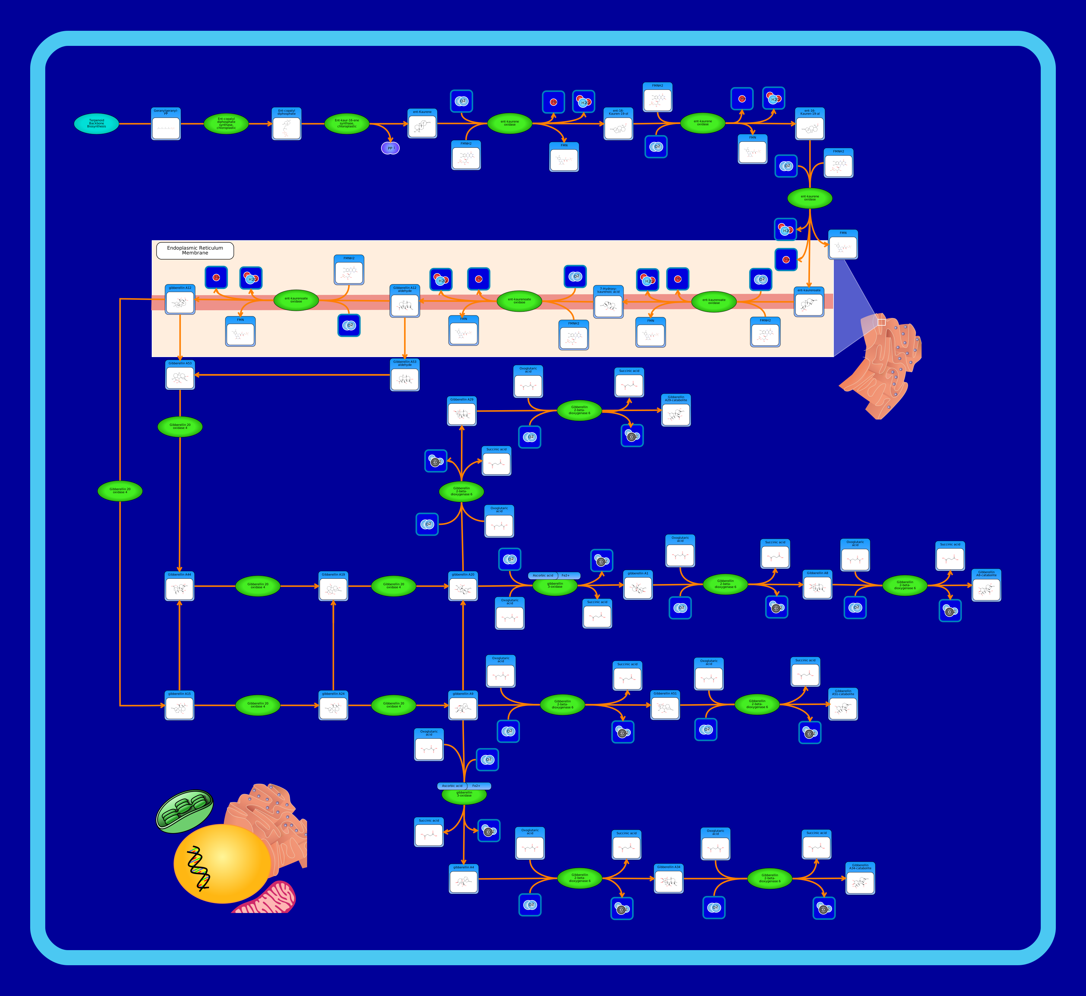 View Pathway
|
Gibberellin and Gibberellin Precursor Biosynthesis PathwayArabidopsis thaliana
A gibberellin is a plant hormone that is necessary for many functions within a plant. Some of these functions include shoot length, cell division, and bolting induction. This pathway shows the biosynthesis of gibberellin precursors, within the larger diterpenoid biosynthesis pathway. It begins with geranygeranyl-PP, synthesized from Terpenoid backbone biosynthesis, using the enzyme ent-copalyl diphosphate synthase to create ent-copalyl diphosphate. From here, ent-copalyl diphosphate uses ent-kaur-16-ene synthase to create ent-kaurene. Ent-kaurene continues this pathway, teaming up with ent-kaurene oxidase to produce ent-16-kauren-19-ol. Ent-kaurene oxidase is used again in combination with ent-16-kauren-19-ol to synthesize ent-16-kauren-19-al. The enzyme ent-kaurene oxidase is used once more, to create ent-kaurenoate. At this point in the pathway, the reactions begin to take place in the endoplasmic reticulum membrane. Here, ent-kaurenoae oxidase converts ent-kaurenoate into 7-hydroxy-kaurenoic acid. This acid then uses ent-kaurenoate oxidase again to become gibberellin A12 aldehyde. Exiting the endoplasmic reticulum membrane, gibberellin A12 uses gibberellin 20 oxidase 4 to produce gibberellin A15. The reactions continue, still using gibberellin 20 oxidase 4 to create gibberellin A24, and subsequently gibberellin A9. After this, gibberellin a9 is converted to gibberellin a51 through the enzyme gibberellin 2-beta-dioxygenase 6. Gibberellin A51 then uses this same enzyme to create a gibberellin A51-catabolite. There are many offshoots in this pathway that are not described in detail, but all assume a similar chain of reactions and ultimately result in the production of different gibberellin catabolites.
|
Metabolite
Metabolic
|
|
|
SMP0121209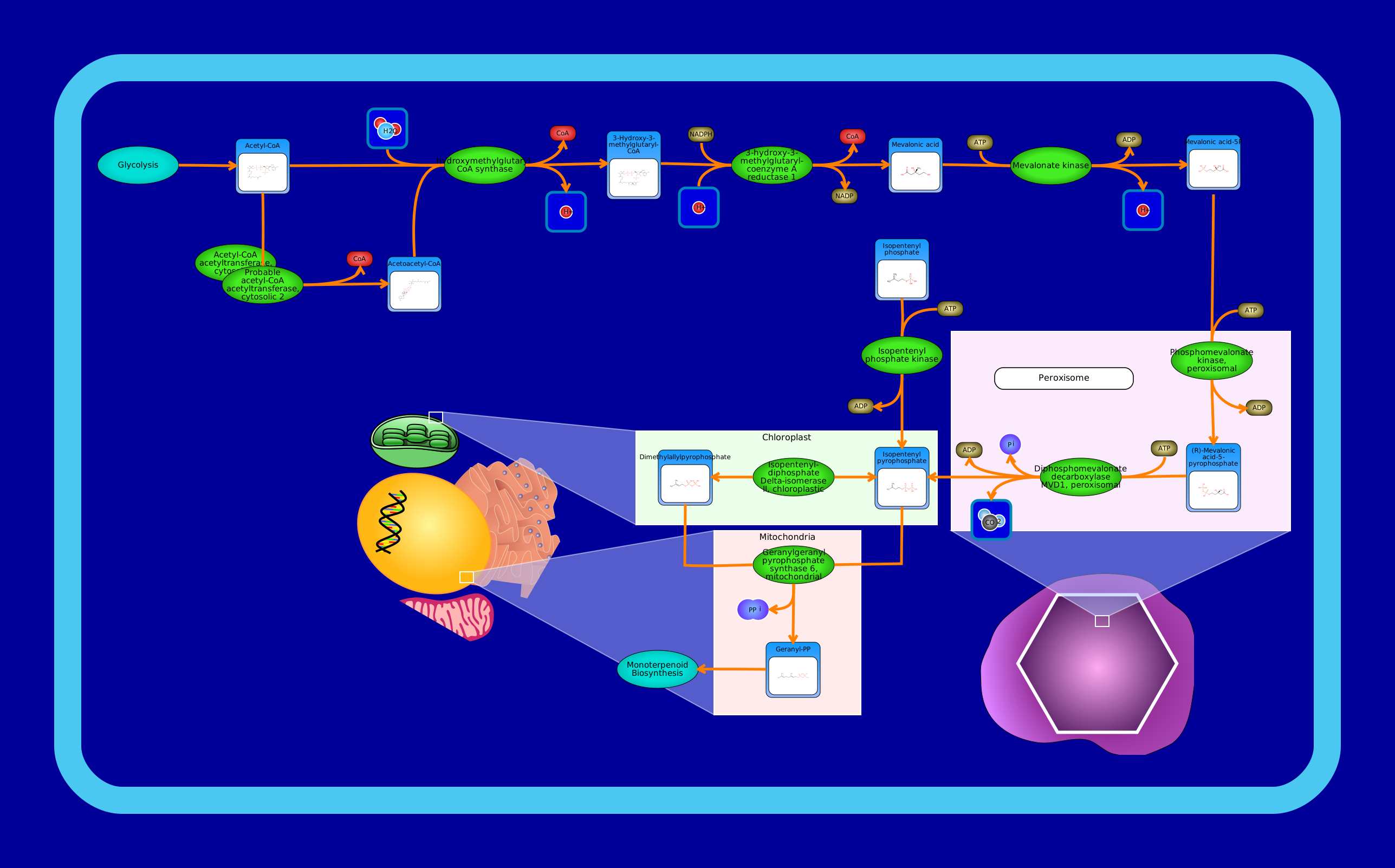 View Pathway
|
Mevalonate PathwayArabidopsis thaliana
The mevalonate pathway, also known as the isoprenoid pathway, plays an essential role in creating the chemicals needed for many plants to function. This pathway, combined with the MEP/DOXP pathway give many plants their scents, such as cinnamon and ginger, and are responsible for the red colour in tomatoes. The pathway begins with acetyl-CoA, having come from the glycolysis pathway. Acetyl-CoA immediately becomes acetoacetyl-CoA through the enzyme acetyl-CoA acetyltransferase 1/2. Combined, acetoacetyl-CoA and acetyl-CoA react with hydroxymethylglutaryl-CoA synthase to create 3-hydroxy-3methylglutaryl-CoA. From here, this compound is catalyzed by 3-hydroxy-3-methylglutaryl-coenzyme A reductase 1 and becomes (R)-mevalonate. Mevalonate is paired with mevalonate kinase to produce mevalonic acid-5P. In turn, mevalonic acid-5P reacts with phosphomevalonate kinase, and entering the peroxisome and becoming (R)-mevalonic acid-5-pyrophosphate. Remaining in the peroxisome, diphosphomevalonate decarboxylase MVD1 is used alongside (R)-mevalonic acid-5-pyrophosphate to create isopentenyl pyrophosphate, bringing the pathway into the chloroplast. Dimethylallylpyrophosphate is produced after isopentenyl pyrophosphate and isopentenyl diphosphate delta-isomerase II team up to catalyze it. Dimethylallylpyrophosphate then joins forces with isopentenyl again, this time adding geranylgeranyl pyrophosphate synthase 6 and moving into the mitochondria to produce geranyl-PP. This is followed by monoterpenoid biosynthesis.
|
Metabolite
Metabolic
|
|
|
SMP0122304 View Pathway
|
Rhamnolipid BiosynthesisPseudomonas aeruginosa
Rhamnolipids (RL) consist of a fatty acyl moiety composed of a 3-(3-hydroxyalkanoyloxy)alkaloid acid (HAA) and a sugar moiety composed of one or two rhamnose sugars. Rhamnolipids function as surfactants and virulence factors and are involved in biofilm formation and cell motility. The rhamnose sugar component is produced via the dTDP-L-rhamnose biosynthetic pathway which forms dTDP-L-rhamnose from glucose 6-phosphate (G6P) in five steps. First, glucose 6-phosphate is converted into glucose 1-phosphate (G1P) via the enzyme phosphoglucomutase (AlgC). Second, glucose 1-phosphate is converted into dTDP-D-glucose via the enzyme glucose-1-phosphate thymidylyltransferase (RmlA). Third, dTDP-D-glucose is converted into dTDP-4-dehydro-6-deoxy-D-glucose via the enzyme dTDP-glucose 4,6-dehydratase (RmlB). Fourth, dTDP-4-dehydro-6-deoxy-D-glucose is converted into dTDP-4-dehydro-L-rhamnose via the enzyme dTDP-4-dehydrorhamnose 3,5-epimerase (RmlC). Fifth, dTDP-4-dehydro-L-rhamnose is converted into dTDP-L-rhamnose via the enzyme dTDP-4-dehydrorhamnose reductase (RmlD). The HAA component is synthesized from 3-hydroxyacyl-[acyl-carrier protein] diverted from fatty acid biosynthesis via the enzyme 3-(3-hydroxydecanoyloxy)decanoate synthase (RhIA). The final step in rhamnolipid biosynthesis is the formation of the glycosidic link between the rhamnose sugar component and the HAA component. This is accomplished by two rhamnosyltransferases (RhlB and RhlC) which catalyze sequential glycosyl transfer reactions to first form mono-rhamnolipids (via RhIB) and then di-rhamnolipids (via RhIC). RHlA, RHlB, and RHlC are associated with the inner membrane.
|
Metabolite
Metabolic
|
||
SMP0030406 View Pathway
|
Androstenedione MetabolismHomo sapiens
Androstenedione is an endogenous weak androgen steroid hormone that is a precursor of testosterone and other androgens, as well as of estrogens like estrone . Its metabolism occurs primarily in the endoplasmic reticulum (membrane-associated enzymes are coloured dark green in the image). Conversion of androstenedione to testosterone requires the enzyme testosterone 17-beta-dehydrogenase 3. Conversion of androstenedione to estrone involves three successive reactions catalyzed by the enzyme aromatase (cytochrome P450 19A1). Androstenedione can also be converted into etiocholanolone glucuronide, androsterone glucuronide, and adrenosterone. The three-reaction subpathway to synthesize etiocholanolone glucuronide begins with the enzyme 3-oxo-5-beta-steroid 4-dehydrogenase catalyzing the conversion of androstenedione to etiocholanedione. This is followed by the conversion of etiocholanedione to etiocholanolone which is catalyzed by aldo-keto reductase family 1 member C4. Lastly, the large membrane-associated multimer UDP-glucuronosyltransferase 1-1 catalyzes the conversion of etiocholanolone to etiocholanolone glucuronide. The three-reaction subpathway to synthesize androsterone glucuronide begins with the conversion of androstenedione to androstanedione via 3-oxo-5-alpha-steroid 4-dehydrogenase 1. Anstrostanedione is then converted into androsterone via aldo-keto reductase family 1 member C4. The last reaction to form androsterone glucuronide is catalyzed by the large multimer UDP-glucuronosyltransferase 1-1. The two-reaction subpathway to synthesize adrenosterone begins in the mitochondrial inner membrane where androstenedione is first converted into 11beta-hydroxyandrost-4-ene-3,17-dione by the enzyme cytochrome P450 11B1. Following transport to the endoplasmic reticulum, 11beta-hydroxyandrost-4-ene-3,17-dione is converted into adrenosterone via corticosteroid 11-beta-dehydrogenase isozyme 1.
|
Metabolite
Metabolic
|
|
|
SMP0063472 View Pathway
|
Vitamin B6 MetabolismArabidopsis thaliana
Vitamin B6 is a water-soluble vitamin essential for all living organisms. It is an important cofactor for enzymatic reactions in over one hundred different cellular reactions and processes. Vitamin B6 exists in different natural forms called vitamers, which are produced by plants, bacteria, and fungi, but not by animals and humans. These vitamers include: pyridoxal (PL), pyridoxine (PN) and pyridoxamine (PM) and their phosphorylated vitamers, PLP, PNP and PMP respectively. Vitamin B6 metabolic pathway was mainly characterized in E. coli, however most organisms, including plants, utilize an alternate pathway. In plants, the various vitamers can be produced via different specific pathways. In A. thaliana, this biosynthetic pathway involves few subpathways, which include: glycolysis, pentose phosphate pathway (PPP), and glyoxylate and dicarboxylate metabolism. Glyceraldehyde 3-phosphate produced by glycolysis and ribulose 5-phosphate produced by PPP are synthesized to pyridoxal 5-phosphate by a synthase. Pyridoxal 5-phosphate is then dephosphorylated to pyridoxal. Pyridoxal, a form of vitamin B6, could act as a precursor for butanoate metabolsim. Moreover, from PPP, 2-Oxo-3-hydroxy-4-phosphobutanoate is produced, this is synthesized to O-phospho-4-hydroxy-L-threonine and then to 4-hydroxy-L-threonine. Pyridoxine could also be produced after a multistep reaction from 4-hydroxy-L-threonine, which is then synthesized to pyridoxal. Glycoaldehyde produced from glyoxylate and dicarboxylate metabolism is converted to pyridoxine. Pyridoxine could also undergo phosphorylation where it is converted to pyridoxine phosphate which is then synthesized to pyridoxal 5-phosphate where the later is dephosphorylated to pyridoxal. Pyridoxal could also be synthesized to pyridoxamine, this that is phosphorylated to pyridoxamin 5-phosphate, which is then synthesized to pyridoxal 5-phosphate.
|
Metabolite
Metabolic
|
|
|
SMP0014205 View Pathway
|
Phosphatidylcholine BiosynthesisArabidopsis thaliana
Phosphatidylcholines (PC) are a class of phospholipids that incorporate a phosphocholine headgroup into a diacylglycerol backbone. They are the most abundant phospholipid in eukaryotic cell membranes and has both structural and signalling roles. In eukaryotes, there exist two phosphatidylcholine biosynthesis pathways: the Kennedy pathway and the methylation pathway. The Kennedy pathway begins with the direct phosphorylation of free choline into phosphocholine followed by conversion into CDP-choline and subsequently phosphatidylcholine. It is the major synthesis route in animals. The methylation pathway involves the 3 successive methylations of phosphoethanolamine to form phosphocholine which is then funnelled into the Kennedy pathway to make phosphatidylcholine. In plants, phosphatidylcholine biosynthesis is implemented using a mix between the two pathways. An alternative of the methylation pathway uses phosphatidylethanolamine as a starting compound, but no enzyme has been found in Arabidopsis to catalyze the first methylation to form phosphatidyl-N-methylethanolamine. Many enzymes involved in this pathway are localized to the cell membrane but are not drawn as such for clarity. Instead, they are indicated with a dark green colour and appear to be free floating in the cytosol. The first reaction of the Kennedy pathway involves the membrane-localized enzyme choline/ethanolamine kinase catalyzing the conversion of choline into phosphocholine. Second, choline-phosphate cytidylyltransferase catalyzes the conversion of phosphocholine to CDP-choline. Last, choline/ethanolaminephosphotransferase, localized to the cell membrane, catalyzes phosphatidylcholine biosynthesis from CDP-choline. It requires either magnesium or manganese ions as cofactors. Note that phosphatidylcholine can be converted to either phosphocholine by a non-specific phospholipase or converted to choline by phospholipase D. Phosphocholine can also be converted to choline via phosphoethanolamine/phosphocholine phosphatase. The methylation pathway begins with serine decarboxylase catalyzing the biosynthesis of ethanolamine from serine. It requires pyridoxal 5'-phosphate as a cofactor. Next, choline/ethanolamine kinase, localized to the cell membrane, catalyzes the conversion of ethanolamine to phosphoethanolamine. Phosphoethanolamine N-methyltransferase (PEAMT), located in the cytosol, then catalyzes three sequential N-methylation steps to convert phosphoethanolamine to phosphocholine. PEAMT uses S-adenosyl-L-methionine as a methyl donor. Phosphocholine then enters the Kennedy pathway. Alternatively, in a subpathway parallel to the Kennedy pathway, phosphoethanolamine can be converted into phosphatidylethanolamine. Phosphatidylethanolamine is also synthesized from phosphatidylserine in the endoplasmic reticulum by phosphatidylserine decarboxylase. Note that phosphatidylethanolamine can be converted to either phosphoethanolamine by a non-specific phospholipase or converted to ethanolamine by phospholipase D. The two methylated intermediates N-methylethanolamine phosphate and N-dimethylethanolamine phosphate can also undergo reactions parallel to the Kennedy pathway to form the methylated intermediates of phosphatidylethanolamine (otherwise catalyzed by phosphatidyl-N-methylethanolamine N-methyltransferase, localized to the endoplasmic reticulum membrane, to form phosphatidylcholine).
|
Metabolite
Metabolic
|
|
|
SMP0012467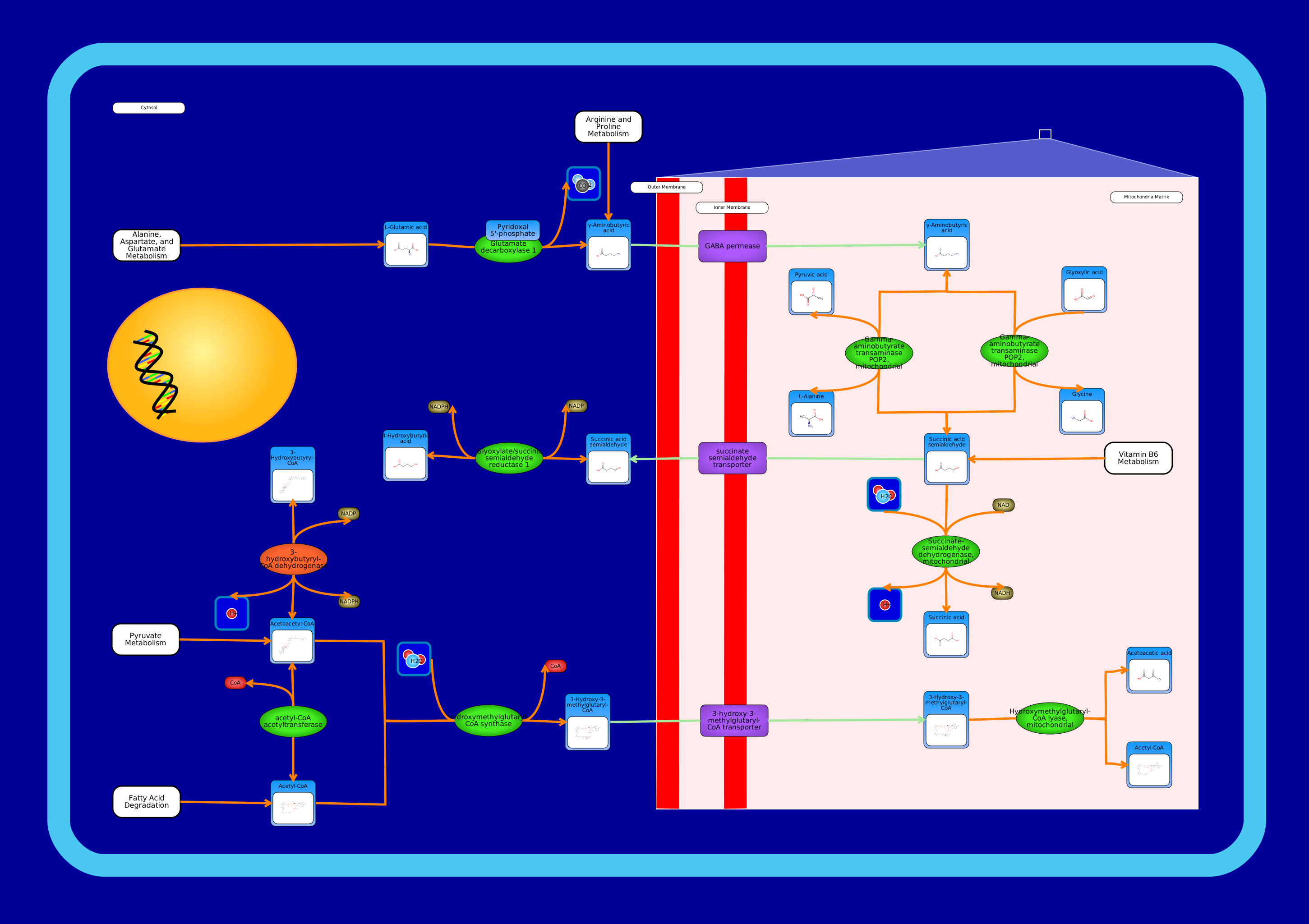 View Pathway
|
Butanoate MetabolismArabidopsis thaliana
Butanoate or butyrate is the traditional name for the conjugate base of butanoic acid (also known as butyric acid). Butanoate metabolism includes L-glutamate degradation into the signal molecule GABA followed by subsequent reactions to make further products. Glutamate decarboxylase is an enzyme in the cytosol that catalyzes the conversion of L-glutamate into 4-aminobutanoate (GABA). It requires pyridoxal 5'-phosphate as a cofactor. This is followed by GABA permease, belonging to the APC Family of transport proteins, transporting GABA from the cytosol into the mitochondria matrix. Next, gamma-aminobutyrate transaminase degrades gamma-amino butyric acid (GABA) into succinate semialdehyde and uses either pyruvate or glyoxylate as an amino-group acceptor. The pyruvate-dependent activity is reversible while the glyoxylate-dependent activity is irreversible. Afterwards, succinate-semialdehyde dehydrogenase oxidizes succinate semialdehyde into succinate. A predicted succinate semialdehyde transporter in the mitochondria inner membrane is theorized to export succinate semialdehyde from the mitochondrial matrix into the cytosol. There, glyoxylate/succinic semialdehyde reductase catalyzes the reversible conversion of succinate semialdehyde into 4-hydroxybutanoate. Butanoate metabolism in Arabidopsis thaliana also includes reactions involving acetyl-CoA and acetoacetyl-CoA. 3-hydroxybutyryl-CoA dehydrogenase is a predicted enzyme (coloured orange in the image) in the cytosol that is theorized to catalyze the reversible conversion of 3-hydroxybutanoyl-CoA into acetoacetyl-CoA. Acetyl-CoA acetyltransferase then catalyzes the reversible conversion of acetoacetyl-CoA into acetyl-CoA. Then, hydroxymethylglutaryl-CoA synthase condenses acetyl-CoA with acetoacetyl-CoA to form 3-hydroxy-3-methylglutaryl-CoA. This is followed by a predicted 3-hydroxy-3-methylglutaryl-CoA transporter localized to the mitochondria inner membrane that is theorized to import 3-hydroxy-3-methylglutaryl-CoA into the mitochondrial matrix from the cytosol. Once there, hydroxymethylglutaryl-CoA lyase catalyzes the synthesis of acetoacetate and acetyl-CoA from 3-hydroxy-3-methylglutaryl-CoA.
|
Metabolite
Metabolic
|
|
|
SMP0012089 View Pathway
|
PhotosynthesisArabidopsis thaliana
Photosynthesis involves the transfer and harvesting of energy from sunlight and the fixation of carbon dioxide into carbohydrates. This process occurs in higher plants, including Arabidopsis thaliana. Oxygenic photosynthesis requires water, which acts as an electron donor molecule. The reactions which involve the trapping of sunlight are known as "light reactions", and result in the production of NADPH, adenosine triphosphate, and molecular oxygen. The "dark reactions" are known as the Calvin cycle, and involve the use of the products of the light reactions to fix carbon dioxide and produce carbohydrates. Photosynthesis begins with photosystem II, located in the thylakoid membrane within chloroplasts, which captures light energy to transfer electrons from water to plastoquinone. This process generates oxygen as well as a proton gradient used to synthesize ATP. The D1/D2 (psbA/psbD) reaction center heterodimer binds P680, the primary electron donor of PSII as well as several subsequent electron acceptors. Next, the cytochrome b6-f complex mediates electron transfer between photosystem II (PSII) and photosystem I (PSI). Plastoquinol shuttles electrons from PSII to cytochrome b6-f complex. Plastocyanin shuttles electrons from cytochrome b6-f complex to PSI. Photosystem I is a plastocyanin-ferredoxin oxidoreductase which uses light energy to transfer an electron from the donor P700 chlorophyll pair to the electron acceptors A0, A1, FX, FA and FB in turn. The function of PSI is to produce the NADPH necessary for the reduction of CO2 in the Calvin-Benson cycle. Finally, the proton gradient allows ATPase to synthesize ATP from ADP. The light-independent Calvin-Benson cycle consist of nine reactions that take place in the chloroplast stroma. Beginning with the enzyme RuBisCO, D-ribulose-1,5-bisphosphate is converted into 3-phosphoglyceric acid. It requires magnesium ion as a cofactor. Next, chloroplastic glyceraldehyde 3-phosphate dehydrogenase catalyzes the conversion of glyceric acid 1,3-biphosphate into D-glyceraldehyde 3-phosphate. Then triose-phosphate isomerase catalyzes the conversion of D-glyceraldehyde 3-phosphate into dihydroxyacetone phosphate. Next, the enzyme fructose-bisphosphate aldolase catalyzes the conversion of dihydroxyacetone phosphate into fructose 1,6-bisphosphate. Then fructose-1,6-bisphosphatase catalyzes the conversion of fructose 1,6-bisphosphate into fructose-6-phosphate. It requires magnesium ion as a cofactor. Next, transketolase catalyzes the conversion of fructose-6-phosphate into xylulose 5-phosphate. It requires a divalent metal cation and thiamine diphosphate as cofactors. Then the enzyme ribulose-phosphate 3-epimerase is catalyzes the interconverson of xylulose 5-phosphate and D-ribulose 5-phosphate. Lastly, phosphoribulokinase catalyzes the conversion of D-ribulose 5-phosphate to regenerate D-ribulose-1,5-bisphosphate. An alternative pathway intersects the Calvin-Benson cycle providing another route to synthesize D-ribulose 5-phosphate and D-xylulose 5-phosphate, which both feed back into the main cycle, from dihydroxyacetone phosphate. This subpathway begins with the predicted enzyme sedoheptulose-1,7-bisphosphate aldolase theorized to catalyze the converson of glycerone phosphate and D-erythrose 4-phosphate into sedoheptulose-1,7-bisphosphate. Next, sedoheptulose-1,7-bisphosphatase catalyzes the conversion of sedoheptulose-1,7-bisphosphate into D-sedoheptulose 7-phosphate. Next, transketolase catalyzes the converson of D-sedoheptulose 7-phosphate into D-ribose 5-phosphate and D-xylulose 5-phosphate (which feeds back into the main cycle). Lastly, ribose-5-phosphate isomerase is the probable enzyme that catalyzes the interconverson of D-ribose 5-phosphate and D-ribulose 5-phosphate. D-ribulose 5-phosphate feeds back into the main cycle.
|
Metabolite
Metabolic
|
|
|
Showing 21 -
30 of 541248 pathways